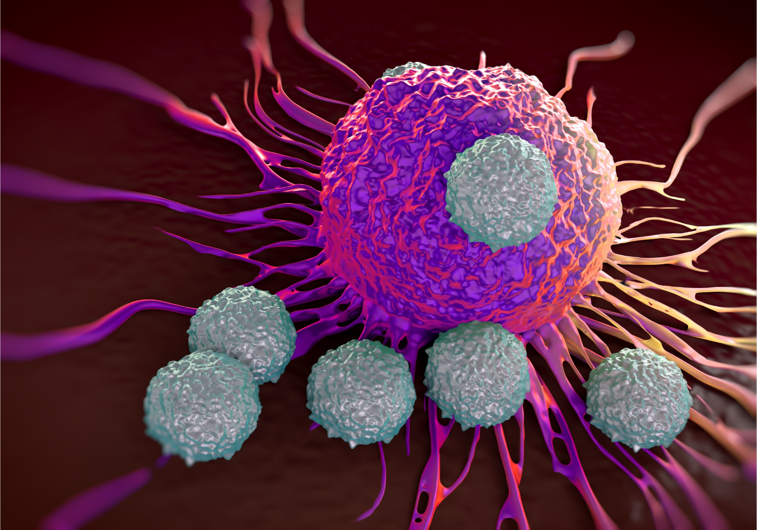
Science Translational Medicine
The Mushett Family Foundation
The New York Academy of Sciences
Help the New York Academy of Sciences bring late-breaking scientific information about the COVID-19 pandemic to global audiences. Please make a tax-deductible gift today.
DONATEPosted June 09, 2017
Presented By
Cancer immunotherapy’s ability to harness the body’s own immune system to target and kill tumor cells has proven to be clinically formidable. Now that checkpoint blockades have been developed to squelch the immune system’s own negative regulatory mechanisms and release cytotoxic T cells from suppression, researchers and clinicians are finding tumor antigens to use as targets and synthesizing optimized receptors to target them. On February 27 and 28, 2017, scientists and physicians convened at the New York Academy of Sciences for Frontiers in Cancer Immunotherapy, a symposium focused on toxicities induced by these therapies, and methods for dealing with the resistance that develops in some populations.
This program was supported in part by educational grants from AstraZeneca, Bristol-Myers Squibb, Genentech, Incyte, and Prometheus Laboratories Inc.
Cancer cells express and display novel antigens that can be recognized and attacked by the immune system. This insight could not be exploited therapeutically, however, until researchers realized that the tumor microenvironment actively suppresses the immune system, in the same manner as successful pathogens. As a result, immune checkpoint blockades—antibodies targeting and neutralizing PD-1, PD-L1, and CTLA-4—have now been used to release cytotoxic T cells from this immunosuppresssion and successfully treat patients with melanoma and leukemia.
But as is often the case with promising new treatments, once the initial euphoria of success fades, issues of toxicity and resistance arise. Various technologies are showing promise to combat these factors, as well as methods for extending the utility of immune checkpoint blockades to solid tumors, which have traditionally been more recalcitrant. The way forward likely includes identifying new antigens in tumors, both those that have never been targeted by immune checkpoint blockade and those that have escaped through by antigen loss; optimizing recombinant and synthetic immune molecules like CARs; and finding biomarkers to better identify patients who might most benefit from immunologically based therapies as well as patients who might most suffer toxic effects from them. But current limitations in the field, notably its reliance on animal models of questionable applicability, must first be overcome.
Speakers tackled these topics from a variety of angles.
Padmanee Sharma’s keynote address focused on MD Anderson’s emphasis on both clinical trials and laboratory research, and how the responses of patients treated with ipilimumab informed their experiments and led them to identify the role of the ICOS pathway in CTLA-4 blockade therapy.
Philip Greenberg discussed identification of WT-1 as a potent tumor antigen and generation of CD8+ T cells that specifically recognize it to treat leukemia patients who relapsed after receiving bone marrow transplants. Nina Bhardwaj explained how the tumor microenvironment promotes immunosuppression by stimulating dendritic cells.
Jedd D. Wolchok noted that combination therapy seems to be more effective, but also more toxic, than monotherapies blocking either PD-1 or CTLA-4 alone. Stephen Baylin said that most cancers contain epigenetic abnormalities, and showed that epigenetic drugs can be used to render tumors more susceptible to immunotherapies. Cornelia Trimble delved into therapeutic vaccination for premalignant HPV disease, and continual refinements to the treatment.
Lisa Coussens stressed that tumor infiltrating lymphocytes resemble cells in their homeostatic tissues more than they do infiltrates in other tumors. Mark Davis talked about technologies that he has developed to synthesize tumor specific T cells for clinical use. Antoni Ribas pointed out that patients’ PD-1 and PDL-1 status do not necessarily predict their susceptibility to PD-1 checkpoint blockade therapy, meaning better biomarkers are desperately needed.
Thomas Gajewski described the myriad tumor and host pathways that shape degrees of immunotherapy resistance, while Andrea Schietinger described mouse studies that have revealed different molecular and epigenetic pathways underlying CD8+ T cell dysfunction and exhaustion in solid tumors, Hiroyoshi Nishikawa discussed the seemingly contradictory roles the transcription factor FoxP3 can play in colorectal cancer, and, Carl June talked about how synthetic biology can be harnessed to engineer tumor-specific CAR cells.
James Yang described his mutation recognition algorithm for identifying tumor neoantigens in silico, Michele Maio reminded the assembled that HLA class I downregulation remains a potent and neglected aspect of tumor resistance to immunotherapy, and Matthew Gubin announced that personalized cancer vaccine can now be generated within eight weeks of a tissue biopsy. Alexander Rudensky talked about Treg cells in the tumor microenvironment; Crystal Mackall warned that antigen escape may become a serious threat to CAR therapies as these therapies move on from leukemia, where CD19 CAR can be thwarted by CD19 loss, into solid tumors; and Kunle Odunsi spoke about tryptophan catabolism as a potential target to alleviate T cell suppression.
Finally, short hot topics talks covered everything from how activation of the brain’s reward center can stimulate the immune system’s cancer fighting abilities to fatty acid metabolism as a regulator of T cell lineage and longevity to the use of a combination of autologous cytokines as a vaccine. The conference ended with a keynote address by Laurence Zitvogel on the vital role that the microbiome plays in regulating immunity, and how it may be harnessed therapeutically.
Keynote Speaker
Padmanee Sharma
The University of Texas MD Anderson Cancer Center
Padmanee Sharma
Immune checkpoint blockades can be used to combat many tumor types because they don’t target tumors, they target the immune system.
A “reverse translation” approach operates by linking clinical trials and lab work, using what works in patients to generate hypotheses to test.
Immune systems display a “yin and yang” effect. Turning one component on triggers the body’s defenses to work to turn it off.
Stimulation through the T cell receptor is insufficient to unleash antitumor immune responses—a failing identified by the first tumor vaccine trials. Researchers learned that costimulation by dendritic cells through CD28 is also required, while at the same time, off signals—such as those mediated by CTLA-4—are needed to maintain immune system homeostasis, as their blocking is necessary to allow for tumor eradication. The first phase I trial done utilizing this insight was of ipilimumab, an antibody that blocks CTLA-4. It was successful in eradicating metastatic melanoma, and its approval in 2011 opened up the field of immune checkpoint therapy. Other drugs blocking other immune checkpoint molecules, notably PD1 and its ligand PD-L1, have followed.
Yet “clinical trials currently outstrip our understanding of the cellular and molecular mechanisms underlying both efficacy and toxicity,” said Padmanee Sharma of the University of Texas MD Anderson Cancer Center. Thus, MD Anderson’s immunotherapy platform takes a “reverse translation” approach linking clinical trials to lab work. Instead of starting with a hypothesis and developing therapies for patients, they use what works in patients to generate hypotheses to test in the lab and identify biomarkers. In this way, her lab worked back from the observation that treatment with ipilimumab before bladder resection acted to enrich T cell infiltration into tumors, and that proteins in the ICOS pathway were highly upregulated in the infiltrating T cells. This surprising finding—ICOS had never before been shown to have antitumor activity although it had been correlated with increased survival in metastatic melanoma—led them to hypothesize that ipilimumab works through the ICOS pathway. Experiments using ICOS knockout mice confirmed that this was in fact the case, suggesting that targeting ICOS as a combination therapy with ipilimumab might be an effective antitumor strategy.
This observation that treatment with ipilimumab before surgery enriched T cell infiltration into tumors led to the idea that ipilimumab might also be harnessed to treat other tumor types with lower mutational loads than those typically seen in bladder cancer and melanoma, even those that lack CTLA-4, for instance prostate cancer. Prostate cancer has very little tumor infiltration, and is thus not a great candidate for immune checkpoint therapy. According to Sharma, however, if treatment with ipilimumab can increase T cell infiltration into prostate cancers and open it up to immunotherapy, doctors might be able to ratchet up life expectancies by years rather than months.
Speakers
Phillip D. Greenberg
University of Washington
Nina Bhardwaj
Mount Sinai School of Medicine
Chimeric antigen receptors can be used to engineer adaptive immune cells and impart specificity to T cells.
The tumor microenvironment is key to modulating immunotherapy.
Orthogonal classes of immunomodulating drugs can be combined synergistically to combat tumors.
Philip Greenberg of the University of Washington went looking for ways to render the immune system effective at fighting tumors by engineering adaptive immune cells. Chimeric antigen receptors (CARs) purport to do just that. By tinkering with the antigen and the T cell receptor, researchers and clinicians can now impart a desired specificity to T cells. On the plus side, since these CARs are engineered, they are not HLA restricted like natural T cell receptors are. But on the minus side, they need a high density of antigen. They have been very effective at fighting leukemias and lymphomas, but not yet proved themselves effective at fighting solid tumors. This lack of a “one size fits all” engineered T cell prompted Greenberg to explore other feats of engineering.
Graft versus host disease (GVHD) is a serious problem for leukemia patients who get bone marrow transplants, but patients who suffer from GVHD have reduced rates of relapse from their cancers. Since GVHD is T cell mediated, Greenberg’s lab hypothesized that giving patients T cells that can specifically target their tumors might prevent GVHD as well as relapse. They settled on using WT-1 as an antigen. WT-1, for Wilms’ Tumor Antigen 1, is highly overexpressed in leukemia cells where it promotes proliferation, oncogenicity, and clinical aggressiveness. Greenberg’s team isolated CD8+ T cells from blood marrow donors and expanded the clones that reacted most highly with WT-1 tumor cells from patients, then treated them with IL-21 to prolong their survival in vivo. When tried in an initial cohort of eleven patients, most patients required only one infusion of these T cells after their bone marrow transplants. The cells were maintained in their bodies for over a year, and they did not relapse. These T cells are currently in clinical trials against high risk acute myeloid leukemia (AML).
But there are caveats. Although this seems to be a promising strategy to prevent a relapse, it cannot treat one—once patients relapsed, the T cells did not persist nor did they prolong survival. Moreover, although the cells clearly remain functional, it is difficult to tell what their phenotype is. It seems that endogenous T cell receptors get downregulated.
Dendritic cells drain from tumors to lymph nodes, where they activate the T cells that then go on to the tumor. This migration requires a number of different dendritic cells to elicit a good immune response. Nina Bhardwaj of Mount Sinai School of Medicine described two proof of principle studies that demonstrate how we might be able to mobilize these different dendritic cells in order to most effectively induce immune responses against tumors.
The first study looked at Flt3 ligand, which triggers the maturation of dendritic cells and thus promotes their antigen presenting capabilities, making them promising as tumor vaccine adjuvants. In this study, Flt3 ligand was given to high risk melanoma patients along with vaccination with NY-ESO-1 protein, an antigen present in many tumor cells but not normal cells. All patients who got the NYESO-1 vaccine developed a T cell response and antibodies countering the antigen, but the response of those who also got Flt3 ligand “was more robust, was detected earlier, and was detected in more subjects,” Bhardwaj said. Flt3 ligand stimulated both DC1 and DC2 subsets and, “surprisingly,” also elicited an increase in B cells.
The second study was of poly-ICLC, a Toll-like receptor 3 agonist. TLR agonists are very good at priming T cells, which they do by inducing HLA expression in dendritic cells to promote antigen presentation. TLR agonists also directly lead to tumor cell death by activating other innate immune cells, making proinflammatory cytokines, and harnessing immunosuppressive Treg cells. Bhardwaj is in the midst of a Phase II study assessing the efficacy of poly-ICLC against head and neck cancer, skin cancer, and sarcoma. Her early, promising data indicated that patients undergo a complete remission on the therapy. Once they go off, however, they often relapse.
Speakers
Jedd D. Wolchok
Memorial Sloan Kettering Cancer Center
Stephen B. Baylin
Johns Hopkins University
Cornelia (Connie) Liu Trimble
Johns Hopkins University
Blocking critical immune checkpoints also releases T cells that have self-reactivity, and could thus induce toxicity.
Epigenetic therapies try to reverse abnormalities that lead to cancer; reversing them can reverse proliferation.
Peripheral vaccination can impact a localized lesion.
CTLA-4 blockade and PD-1 blockade represent the two most developed immune checkpoints, and as such formed the primary focus for the researchers in attendance. Each have demonstrated efficacy, but both elicit toxicity, with PD-1 blockade eliciting a higher grade, but less frequent, toxicity than CTLA-4. In combination, the two blockades are more effective than either therapy alone, but also more toxic. Interestingly, this dramatic jump in efficacy occurred in only patients whose tumors expressed low levels of PD-1. Jedd Wolchok of Memorial Sloan Kettering Cancer Center speculated that this expression level could be used as a biomarker to ensure that the therapy is used only on those who would benefit most from the combination therapy, and to spare those who would suffer most.
The central tenet of checkpoint blockade therapy is that the best candidates are those whose tumors exhibit a high mutational load and a pretreatment immune response. In order to elicit an immune response in tumors that don’t already do it on their own, Wolchok’s lab uses an oncolytic virus—Newcastle Disease Virus—to generate systemic inflammation, rendering a recalcitrant tumor susceptible to checkpoint blockade therapy. When combined with CTLA-4 blockade, injection of this virus in one tumor can cause T cells to attack distant tumors in mice. Eliminating myeloid-derived suppressor cells from the tumor microenvironment is another strategy his lab is exploring to render tumors more sensitive to immunologically-based therapeutics.
Studies have shown that epigenetic abnormalities—primarily hypermethylation of the CpG rich regions in the promoters of tumor suppressor genes, leading to their downregulation—are present in every cancer. Epigenetic drugs, like histone deacetylase inhibitors and DNA demethylases, thus have as their goal nothing less than reprogramming the entire tumor genome. They do not target specific genes or proteins; they target whole pathways. Epigenetic therapies can synergize with immune based therapies by impacting tumor antigens as well as the chromosomal processing of MHC molecules.
Traditionally, methylation acquired over the course of oncogenesis—for instance of tumor suppressors—was thought to be the most important target of epigenetic drugs. But constitutive methylation, such as that of the repeated viral sequences in our genomes, is important as well. This was demonstrated amply by work showing that a DNA-demethylating agent known to be effective at combating colorectal cancer cells does so not by actively interfering with the methylation of tumorigenic DNA sequences, but rather by inducing double stranded RNAs from endogenous retroviral elements to “trick” colorectal cancer initiating cells into acting like they are experiencing a viral infection.
“This is a good story,” began Cornelia Trimble of Johns Hopkins University, “and I’m a clinician.”
Infection with human papillomavirus can cause cervical cancer. The viral genome integrates into the host genome and directs it to produce two viral proteins, E6 and E7, which are both necessary but insufficient for the cancer to take hold and stay established. They are thus very good target antigens.
Full blown cervical cancers are often preceded by cervical intraepithelial neoplasia (CIN). Some CINs spontaneously regress and don’t become cancerous, but since there is no way of telling in advance which those might be, the standard of care for CIN is surgical resection—which can adversely affect the woman’s reproductive capacity. Trimble vaccinated CIN patients with E6 and E7 in hopes of generating an immune response that would take care of their lesions. They received this treatment before surgery, with the hope that one day it might allow other patients in similar circumstances to forego surgery altogether. She found that about 80% of vaccinated patients experienced histopathological regression to a normal phenotype and viral clearance; 50% of patients who received a mock injection showed these effects. The vaccination was able to elicit E6 and E7 specific CD8+ T cells that remained in normal tissue and in peripheral blood. This is essential for viral clearance, which is in turn essential for preventing recurrent disease. Her study demonstrated that peripheral vaccination can elicit an adaptive immune response that acts on a localized lesion, in this case in the cervix.
In the cervix, T cells congregate immediately outside of the cancerous lesion. They can’t get in because a common mechanism of immune evasion utilized by solid tumors is creation of a slippery barrier outside their neovasculature—they downregulate the expression of adhesion molecules. Trimble thus noted that future immune-based therapeutic strategies must activate the lesion’s epithelium.
Speakers
Lisa Coussens
Oregon Health Sciences University
Mark Davis
Stanford University
Antoni Ribas
University of California Los Angeles
Tumor infiltrating lymphocytes look like T cells in their respective homeostatic tissues, not like T cells in other tumors.
Tumor infiltrating lymphocytes have a higher degree of clonal expansion than T cells in normal tissue.
Myeloid cells, like macrophages, are often associated with more aggressive disease. Tumors subvert their role in tissue remodeling, normally utilized in the context of wound healing, to customize the tumor microenvironment. This makes myeloid cells attractive as druggable targets.
In addition to their role in tumor progression, myeloid cells regulate the functional capacity of CD8+ T cells to interact in antigen-specific ways. It is this regulation of the T cell response, rather than their tissue remodeling abilities, that seems to be their most important role in tumorigenesis. Since tumor infiltrates look like T cells from homeostatic tissues more than they look like T cells from other tumors, the tissue-based rules are the ones to bear in mind in any attempts to reprogram the immune system to fight cancer. It is essential to target metastases as well as primary tumors—but these tumors may be very different from one another. Tissue specificity matters.
No one with mesothelioma survives for even five years; palliative care is the only option that comes with the diagnosis. At Oregon Health Sciences University, Lisa Coussens is working with six different mouse models of mesothelioma with de novo metastases from the intestine to the lung. The tumors are chemoresistant. But when CSF1R inhibitors are used to suppress macrophage presence in the tumors, chemotherapy can induce a CD8+ T cell infiltration into the primary tumor. These T cells reduce growth of the primary tumor—but since they are not recruited to the metastases in the lungs, they do not impact the survival of the mice. The same therapy doesn’t work on the same tumor cells, because they exist in different microenvironments.
Coussen’s bottom line summary is that to be clinically effective, we must consider where in the body the metastases occur. And identifying biomarkers that can act as predictive signatures will be crucial, in order to determine how each patient will respond, and to measure whether a patient is even, in fact, responding.
Meaningful findings in human immunity, and cancer, have lagged behind those in mouse models. Mark Davis of Stanford University thinks that this is due to a lack of technologies and infrastructure for studying human samples, an issue he’s working to address.
One new technique he suggests is a low cost, high throughput way of looking at the T-cell receptor sequence of a single cell and linking it to functionality, since T cells bearing identical receptors can still be functionally distinct if they mature along different lineages. RNA sequencing (RNA-Seq) was not a viable strategy to achieve this goal, since T cells possess much less RNA than other cells. By using cell sorting and deep sequencing of both the TCRα and TCRβ genes in individual cells, Davis’ technique can determine the antigen specificity of successful T cell clones, discovering antigens that can then be targeted therapeutically.
A second innovation is an iteration of the classic yeast two hybrid method that he has modified as a tool for antigen discovery. In this variant, a yeast library of MHC molecules linked to randomized peptides is screened for reactivity with T cells. The hits generated by this technique included unique endogenous antigens as well as neoantigens specific for tumor infiltrating lymphocytes, suggesting that it can be used to identify neoantigens in a wide range of tumors.
By identifying the specificity of TCR repertoires one cell at a time, Davis proposes a “TCR-omics.” These TCR sequences can be used for things like determining which regions are most important for T cell specificity and reading which pathogens a patient has been exposed to.
Programmed Death Ligand 1—PD-L1—has been intuitively used as a biomarker to determine which patients will respond to PD-1 blockade therapy. But PD-L1 can be constitutively expressed by tumor cells, or it can be induced in tumor cells, or it can be induced in normal cells. If a patient is PDL-1 positive but the tumor has no T cell infiltration, PD-1 therapy won’t work; conversely, if a tumor is PD-L1 negative because it has no T cell infiltration, perhaps an immune response would instigate such infiltration, rendering that tumor susceptible to PD-1 therapy. Obviously, PD-L1 is “an imperfect biomarker,” said Antoni Ribas.
Interferon-γ turns on PD-L1 in tumor cells by signaling through the JAK/STAT pathway. Tumors with high mutational loads generally do not respond to PD-1 blockade; this might be because it is beneficial to them to be impervious to IFN-γ signaling. “If tumors are not using PDL-1 to protect themselves,” Ribas asked, “why bother blocking it?”
Speakers
Thomas Gajewski
University of Chicago
Andrea Schietinger
Memorial Sloan Kettering Cancer Center
Hiroyoshi Nishikawa
National Cancer Center Hospital, Japan
Carl June
University of Pennsylvania
T cell inhibitory pathways are not set up by the tumor; they are intrinsic immune system regulatory loops.
Self-tolerance is essential to prevent autoimmunity, but is an obstacle to immune therapy.
Patients that have circulating, proliferating CAR T cells qualify as transgenic humans.
Cancer immunotherapy relies upon a tumor microenvironment that is inflamed with CD8+ T cells at baseline. The bulk of checkpoint blockade therapies target T cell inhibitory pathways that are not set up by the tumor; they are intrinsic immune system regulatory loops. In addition to the commonly used immune checkpoint molecules PD-1, PDL-1, and CTLA-4, Thomas Gajewski's lab at the University of Chicago has found that the transcription factor Egr-2 can also act as an immunosuppressor in the tumor microenvironment. Egr-2 is involved in T cell anergy, wherein the T cell receptor is stimulated without concomitant costimulation, rendering the T cell non-responsive, and in fact recalcitrant, to future activation. These dysfunctional T cells are stimulated by antigen and proliferate, but then undergo apoptosis before they kill their tumor. Immunotherapy that alleviates checkpoints and blockades can rescue their functionality.
By using mouse models to investigate the differences between CD8+ T cells that respond to tumor antigens and those that don't, Andrea Schietinger of Memorial Sloan Kettering Cancer Center found out that not all dysfunctional T cells are created equal. Her studies began with naïve T cells, activated by incubation in a dish with Listeria. Upon differentiating into effector cells, they underwent extensive chromatin remodeling; then as they became memory cells, the epigenetic modification slowed down. The epigenomes of the earlier stage cells were more plastic, rendering those cells reprogrammable.
Like stimulation with Listeria and other microbes, stimulation with tumor antigens also alters chromatin states in T cells. When Schietinger activated naïve T cells by injecting them into mice and then inducing tumors in the mice she witnessed a similar phenomenon to what transpired in her dishes: for about a week the T cell chromatin is accessible, creating a plasticity that is amenable to therapeutic intervention. Past that point, however, the chromatin remodels again and the T cells become fixed in their inactive state. These T cells are activated by the tumor, but since they express PD-1, they don't move against it.
Using computational biology to assess what drives this switch, transcription factor footprint analysis revealed that the first wave of chromatin remodeling during tumorigenesis is accompanied by an upregulation of NFAT target genes in the T cells; during the latter wave, seven days later, genes controlled by the TCF/LEF signaling pathway are downregulated.
The cancer immunoediting hypothesis states that cancers select for and promote immunosuppressive molecules, cells, and behaviors, like FOXP3+ CD4+ T regulatory cells. Infiltration of these Treg cell into tumors is generally associated with poor clinical outcomes in different types of cancers-but in colorectal cancers, FOXP3(+) T cell infiltration was linked to better prognosis in some studies. Approximately 20–30% of colorectal cancers are like melanoma, where FOXP3+ T cells are immunosuppressive and associated with a poor prognosis. But that leaves the other 70–80% of colorectal cancers in which FOXP3 is linked to better survival.
It turns out that there are two variants of FOXP3+ T cells in colorectal tumors. Cells that express high levels of the transcription factor are, in fact, immunosuppressive, as has traditionally been found. But some colorectal tumors also harbor T cells that express lower levels of FOXP3. Not only are these cells not immunosuppressive, they actually secrete inflammatory cytokines. It is these T cells—the ones that express low levels of FOXP3—that are associated with the better prognosis of the colorectal cancers that they infiltrate.
Synthetic biology is a new frontier in medicine, offering the opportunity to generate T cells through the use of either CAR cells or transgenic T cell receptors that are more effective than those that have already evolved.
CARs—chimeric antigen receptors—were first conceived in the early 1990s. They are very attractive as therapeutics because they allow for T cell specificity to be targeted and controlled; they do not require MHC expression and identity or costimulation like endogenous T cell receptors do, and they can circumvent T cell dysfunction, suppression, and exhaustion. CARs went through their first trials in 2010, against B cell leukemia. They registered very high response rates, and researchers found that although they induce B cell aplasia—unable to distinguish between healthy B and cancerous ones, these CD19 specific CAR cells kill them all—that can easily be rescued with intravenous immunoglobulin replacement therapy.
In nonresponding patients, these CD19 CAR T cells do not expand. Engineered cells are designed to act as living products within the patient; unlike all other previous classes of drugs, like small molecules or proteins that are inert, living cells can sense their environment and respond to it accordingly. Although this makes them more challenging to generate and control, it is also the quality that makes them so revolutionary.
Speakers
James C. Yang
National Cancer Institute, U.S. National Institutes of Health
Michele Maio
University Hospital, Sienna
Matthew Gubin
Washington University
Patients refractory to PD-1 blockade can still respond to TIL therapy.
Epigenetic genes can be used to enhance the immunogenicity of tumor cells by impacting immune pathway genes.
In mice, personalized vaccines raised against neoantigens have comparable efficacy to checkpoint blockades.
James Yang of the National Cancer Institute uses genetics to determine therapeutic choices—specifically, to determine which antigens T cell therapies should target. T cell therapies with tumor infiltrating lymphocytes (TILs) are thus far the best treatment for metastatic cancers. Activating the TILs in vitro is ideal, since this separates their activation from the immunosuppressive treatment the patient must undergo for the treatment to be effective. This immunosuppression is required to remove PD-1, a very potent force in the tumor microenvironment. Interestingly, patients who do not respond to PD-1 blockade can still respond to TIL therapy. Yang hypothesized that perhaps part of the reason checkpoint inhibition doesn’t work for them is because they have an insufficient T cell repertoire. This idea was borne out by his observation that tumors with mismatch repair deficiencies, which therefore have a large number of somatic mutations, are hypersensitive to immune checkpoint blockade.
Yang realized that lots of somatic mutations in a tumor means lots of targetable neoantigens—so he devised a way to find them. After whole exome sequencing of a tumor, he identified all of the potential 9- and 10-mers that contain a point mutation and could thus potentially be tumor specific neoantigens. Then he used in silico methods to rank them for predicted peptide-MHC binding. He synthesized the top ten MHC-binding candidate peptides and then screened them to see which actually bound to TIL. These neoantigens have a low potential for autoimmunity and are not at risk of undergoing central thymic tolerance; however, they are entirely patient specific, which is not optimal. Among the hundreds of cells Yang’s team has screened and mutations they’ve found, only one protein has shown up twice as a neoantigen: mutant KRAS. Still, Yang said it’s possible to make “avatars” to represent the tumor and screen for (or generate) T cells against the best antigen using autologous antigen presenting cells loaded with the top neoantigens identified with this mutation recognition technique. The process would take about six weeks and, in theory, could be used to target any type of cancer.
“We don’t always know what we’re targeting, if it’s a driver mutation,” he said. “But if it’s expressed in the tumor and elicits a T cell response, we don’t care.”
A neglected aspect of cancer resistance to immunotherapy is the downregulation of HLA class I molecules by tumor cells, an actually quite common phenomenon. No matter how much we improve the efficacy of T cells, Michele Maio of the University Hospital at Sienna, Italy insists, we need to deal with HLA downregulation if we want to achieve effective therapeutics. Loss of HLA genes is often a hallmark of cancerous lesions that regress after treatment.
In his view, epigenetics provides one of the best ways to combat resistance to immunotherapy, which can also be mediated by the lack of tumor infiltration by TILs. Epigenetic drugs, notably histone deacetylase inhibitors and DNA methyltransferase inhibitors, can be used to alter the expression of immune genes in both T cells and tumor cells. For instance, treatment with DHA has been shown to upregulate the expression of cancer testis antigen and other HLA class I antigens in different tumors, which successfully activated T cells and rendered the tumors susceptible to immune checkpoint blockade therapies. These drugs have been used to treat leukemias for about twenty years to reverse the epigenetic silencing in those cancers. The newer, second generation of drugs is more stable and easier to inject.
Cancers initially develop in immunocompetent hosts. As such, once tumors are clinically detectable they have undergone a fair degree of “editing” by the immune system; they have probably already shed their most immunogenic antigens in their relentless quest to grow and grow. Not much is known about the antigens expressed in nascent tumor cells, but based on this reasoning they may be highly immunogenic. Immunodeficient Rag2(-/-) mice make tumors whose cells phenotypically resemble nascent primary tumor cells, and can thus be used as models for tumor cells “unedited” by the immune system. Indeed, they express highly antigenic mutant proteins.
Checkpoint blockades against CTLA-4 and PD-1 have been enormous boons to patients suffering from a number of different types of cancers. The blockades work by releasing T cells from suppression and allowing them to target tumors. But not much is known about which tumor antigens these reactivated T cells are targeting, and more to the point, if these antigens might be viable candidates for generating tumor-specific vaccines. Matthew Gubin, of the Schreiber lab at Washington University, used genomics and bioinformatics approaches to identify tumor-specific mutant proteins targeted by T cells after anti-PD-1 and/or anti-CTLA-4 treatment in mice. Most of the mutations they identified were passenger, versus driver, mutations; even if they are repaired, the tumor still thrives. Gubin’s team then used the identified antigens to make personalized cancer vaccines—a process that took only eight weeks—and found that the vaccines were as effective as the checkpoint therapy. Since they are patient-specific they induce less toxicity; but they only seem to be effective on small tumors, and only early on. Tumors that were larger or further along in development required a combination of the vaccine plus checkpoint therapy.
Speakers
Alexander Rudensky
Memorial Sloan Kettering Cancer Center
Kunle Odunsi
Roswell Park Cancer Institute
Crystal Mackall
Stanford University
The tumor microenvironment promotes the differentiation of Treg cells.
CD19 loss as a mechanism for B cell ALL to escape from CD19 CAR therapy may be “a canary in the coal mine” as CAR molecules become therapeutics for solid tumors.
Checkpoint blockades have been amazingly successful at treating melanoma, non-small cell lung cancer, kidney cancer, bladder cancer, head and neck cancers, and Hodgkin’s lymphoma. But they have not been especially successful in treating solid tumors, like sarcomas and carcinomas. Alexander Rudensky’s lab at Memorial Sloan Kettering Cancer Center explores another mechanism by which tumors suppress the immune system—by recruiting Treg cells—to see how this type of immunosuppression might be reversed therapeutically.
Treg cells are prominent in the microenvironment of solid tumors, and are thought to contribute to tumor progression. In mouse models of breast carcinomas, Rudensky showed that ablating these Treg cells induces apoptosis in the tumor cells and slows tumor progression to a degree comparable to that achieved by checkpoint blockade. This antitumor activity is dependent on CD4+ T cells but not CD8+ T cells, whereas CTLA-4 and PD-1 checkpoint blockade requires CD8+ T cells but not CD4+ T cells.
Rudensky’s lab followed up these findings in mice with studies of human breast carcinomas. They found that Treg cells in the tumor microenvironment were potently immunosuppressive and resembled Treg cells from normal breast tissue but not from peripheral blood. They had upregulated levels of the chemokine receptor CCR8, which may therefore be a good therapeutic target. Treg cells in lung tumors had upregulated levels of amphiregulin, an EGFR-ligand that is important in tissue modeling and repair during inflammation and injury. It thus likewise may provide a druggable target.
CD19 CARs with a CD28 costimulatory domain have provided a major advance in the treatment of childhood leukemia. Still, there is only about a 60% response rate. CD19 seemed to be as optimal a therapeutic target as could exist, since B cell leukemias are defined by its expression. But remarkably, when exposed to immunotherapy, B cell acute lymphoblastic leukemias (ALL) downregulate its expression. This loss of CD19 is the most common cause of relapse after CD19 CAR therapy. The leukemias perform this antigen loss escape by losing either exon 2 of CD19, in the extracellular region, or exons 5 and 6, in the transmembrane domain. Either way, the mutant CD19 can no longer bind the CAR. Alternatively, cells switch from making CD19 to making CD11, in effect changing from ALL to AML. Once patients lose CD19, they can be treated with CARs targeting CD22. But although naïve patients treated with CD22 CARs do well, once patients have lost CD19 they tend to lose CD22 pretty quickly as well.
Crystal Mackall of Stanford University warned that CD19 loss as a mechanism for B cell ALL to escape from CD19 CAR therapy may be “a canary in the coal mine.” In attempting CAR therapy as a treatment option for solid tumors, antigen loss escape may become a problem even sooner than it did in leukemia, since the requirement for specific antigens in solid tumors isn’t as rigid.
Ovarian cancer develops resistance to chemotherapy very quickly, after a good initial response to front line therapies. Only about 10-12% percent of patients respond to immune checkpoint inhibitors. Women with more highly inflamed tumors—those with more TIL—tend to fare better. Thus Kunle Odunsi of Roswell Park Cancer Institute aimed to alleviate T cell suppression to induce more TILs.
Tryptophan catabolism is a pivotal and underappreciated regulator of innate and adaptive immunity. In the ovarian cancer microenvironment, T cells sense low levels of tryptophan through high levels of uncharged tRNA, which activate the IDO pathway to achieve immunosuppression through T cell cycle arrest, differentiation along the Treg cell lineage, and activation of resting Treg cells. Tumoral IDO1 expression inversely impacts TIL infiltration and patient survival after tumor resection.
With these findings in hand, Odunsi sought to determine if pharmacological inhibition might be a viable strategy for overcoming IDO1 mediated immune suppression and recruiting TILs to ovarian tumors. IDO1 blockade unmasks high avidity polyfunctional TILs isolated from ovarian cancer patients, with a three to five-fold increase in NY-ESO-1 specific response. He is now involved in two clinical trials examining the impact of IDO1 inhibition on ovarian cancer, one of which is designed to see if it will potentiate the response to a vaccine generated against NY-ESO-1. There are also trials underway looking at the effect of combining IDO-1 inhibition with checkpoint blockade in non-small cell lung cancer and metastatic melanoma.
Speakers
Tamar Ben-Shaanan
Technion, Israel Institute of Technology
Lu Huang
The University of Texas M.D. Anderson Cancer Center
Fred T. Valentine
New York University School of Medicine
Roddy O'Connor
University of Pennsylvania
Highlights
Activation of the brain’s reward region enhances immunity.
T cells stimulated by CARs with a CD28 signaling domain become effector cells, while T cells stimulated by CARS with a 4-1BB signaling domain become memory cells.
At Technion, Tamar Ben-Shaanan’s work expands on a finding made twenty-eight years ago that group therapy sessions increased the survival of breast cancer patients. She found that, in mice at least, this placebo effect is mediated by the ventral tegmental area (VTA), a key component of the brain’s reward system that is activated by pleasant activities “like eating and mating,” Ben-Shaanan said.
When she used Designer Receptors Exclusively Activated by Designer Drugs—known as DREADDS, ironically in this case, because they elicited pleasurable feelings—to activate dopaminergic neurons in the mouse VTA, she found a strengthened immune response in the mice. The activity of monocytes, macrophages, and T cells were all increased, and tumors shrank. It became apparent that this effect was mediated by the sympathetic nervous system, known for its role in the fight or flight response to stressful or emergent situations, because when the system was ablated with a toxin the tumors didn’t shrink.
To determine which genes mediate resistance to T cell-mediated tumor cytotoxicity, Lu Huang of the University of Texas M.D. Anderson Cancer Center and colleagues conducted a high-throughput ORF screen to transduce individual gene ORFs into tumor cells in order to observe how the transduction would alter sensitivity of tumor cells to killing by autologous TILs. One of the candidates revealed by the screen was an RNA-binding protein capable of binding to 3' UTRs to destabilize mRNAs.
To confirm this finding, Huang overexpressed this protein in patient-derived melanoma cells, finding that this overexpression does decrease the cells' sensitivity to T cell-mediated cytotoxicity, and conversely, that inhibition of this protein in tumor cells sensitizes them to T cell cytotoxicity. Importantly, overexpression of this protein in melanoma cells was found to dramatically inhibit IFNγ release from autologous TILs, indicating that it blocks T cell recognition of tumor cells. Mechanistically, this protein diminishes surface expression and mRNA levels of HLA-A2 by binding to 3' UTR of HLA-A2 mRNA and destabilizing the mRNA. At the same time, other molecules involved in antigen processing and presentation machinery were also downregulated by the overexpression. Thus, this RNA-binding protein impairs antigen processing and presentation in tumor cells and thus diminishes their recognition and destruction by T cells. As final proof of its importance, the mRNA expression of this gene was found to be higher in non-responders than in responders to PD-1 blockade therapy.
T cell activation by antigens is a very energetically costly proposition, fueled—like many cellular processes—by glucose. In elucidating the ways that different CAR molecules effect their therapeutic work, Roddy O’Connor of the University of Pennsylvania has also shown how different subsets of T cells generate their energy.
Different CAR molecules, especially when they incorporate disparate intracellular signaling domains, have enormous clinical promise. Yet because there is not a functional physiological in vitro model to assess, compare, and contrast the impact of different CAR designs, it has been difficult to generate the optimal receptors to most effectively combat each cancer. O’Connor used mRNA electroporation to express CARs in over 90% of the T cells in a population in vitro without concomitant activation through the endogenous T cell receptor. He found that the CAR signaling domains reprogrammed T cell metabolism, modified T cell bioenergetics and mitochondrial biogenesis, and in this way prevented T cell exhaustion and promoted differentiation along different T cell lineages.
CD28 CAR T cells exhibited enhanced aerobic glycolysis and matured into relatively short lived effector T cells. 4-1BB CAR T cells, in contrast, had enhanced oxidative breakdown of fatty acids, respiratory capacity, and mitochondrial biogenesis; they differentiated into long lived memory T cells.
O’Connor speculated that this knowledge could aid in the design of CAR cells to achieve specific desired functions. For instance, CAR T cells with extensive off tumor targets that might therefore induce a high level of toxicity can purposefully be made to become only short lived effector cells. Moreover, he suggests that a mixture of CD28 like and 4-1BB like CAR T cells might be a better therapy than either one alone, as the combination will provide a suite of T cell types more akin to a natural immune response.
“This is really a vaccine talk,” started Fred Valentine of NYU School of Medicine, “even though we’re using cytokines.”
A cytokine storm can be fatal, but it underscores the point that cytokines work in concert; their effects cannot be isolated. Valentine hypothesized that this full complement of cytokines might mimic the environment of an appropriate immune response, and thus might be used almost like a vaccine: to induce such a response. He stimulated human PBMCs ex vivo with a microbial antigen to generate these cytokines—autologous cytokines being “the only ethical way to do it” because of the risk of infection—and injected the secreted molecules into the lesions of patients with metastatic melanoma.
About 70% of patients developed dense lymphocyte infiltrates—even in nodules that did not receive cytokine injections. A common pattern observed in the patient population was for new nodules to appear while other never-injected nodules in the same patient were regressing. Thirty-eight percent of patients had complete regressions, including a few with stage IV-M1, M2, M3 disease; the median duration of these complete regressions was almost five years. Many other patients continued to survive for several years with their melanoma. Twenty-three percent of patients were still disease free three years after treatment; twenty percent of patients were still disease free after five years.
This immune response was due primarily to the infiltration of CD8+ T cells, and to a lesser extent, CD4+ T cells. NK cells are not involved. Thus, the observed cytotoxicity is MHC-restricted. Valentine’s group could not conclude if the cytokine generated immune response was an enhancement of previously undetectable responses against dominant epitopes or a genuinely new immune response against non-dominant epitopes.
Speaker
Laurence Zitvogel
Institut National de la Santé et Recherche Médicale
Highlights
Administration of broad spectrum antibiotics before treatment abrogates the efficacy of immune checkpoint therapies.
Manipulating the composition of gut microbiota shows potential as a therapeutic avenue exploitable to treat cancer.
One way that cyclophosphamide kills cancer cells is by inducing an anti-tumor immune response. In 2013, Laurence Zitvogel showed that it accomplishes this partly by altering the composition of gut microbiota—about a week after treatment—and causing certain species of Gram+ bacteria—specifically L. johnsonii and E. hirae—to migrate to the spleen. Once there, these bugs induce the differentiation of naïve CD4+ cells into Th1 memory cells and Th17 helper cells. Some of them even become pathogenic Th17 cells, which are essential in mediating cyclophosphamide’s anticancer immune response.
Another cancer drug that more obviously marshals the immune system to attack cancer cells is ipilimumab, an antibody that targets CTLA-4 and thus releases T cells from its suppressive effects. What was not obvious until Zitvogel’s research illustrated it, though, is that this CTLA-4 blockade depends on the microbiome. One injection of the CTLA-4 blockade induces the accumulation of B. fragilis in the small intestine of mice even as it depletes many other Bacteroidales and Burkholderiales species. In both mouse models and actual people with metastatic melanoma, T cell responses specific for B. fragilis and B. thetaiotaomicron were associated with the efficacy of the CTLA-4 blockade; in fact, the bug turned out to be essential for the blockade to be at all effective. Ipilimumab increases the population of immunogenic B. fragilis in the gut, which in turn acts to reinforce its anticancer activity.
The impact of the gut microbiota on the efficacy of the treatment has also been recently extended to the PD-1/PDL-1 blockade in mice and humans (work in progress, AACR2017).
The gut microbiome has at this point been implicated in almost every physiological process, and exhibits an incredibly intertwined relationship with the immune system. Antibiotics that interfere with the microbiome have been shown to also interfere in the activity of chemotherapies. This work shows that sculpting and harnessing the microbiome may also augment the activity of certain classes of drugs.
How useful and relevant are animal models—especially those in which tumors are induced by a viral antigen—in the context of cancers and immunotherapies?
How can we determine when it is optimal to use systemic versus intratumoral injections?
Are Treg cells the cause or effect of the tumor microenvironment’s immunosuppressive properties?
How do epigenetic therapies affect memory T cells?
Is there a difference between T cell dysfunction and T cell exhaustion? If so, what is it?
Should CARs target one antigen at a time, or multiple antigens simultaneously, to best avoid tumor escape by antigen loss?
Why can TILs from a progressing nodule kill autologous melanoma cells ex vivo but not in the tumor?