Getting to the Roots of the ‘Shaking Palsy’
Researchers have learned a lot about the debilitating condition that we now call Parkinson’s Disease over the past 200 years, though some are still stumped by a fascinating neuroscientific question.
Published November 1, 2001
By Fred Moreno, Dan Van Atta, Jill Stolarik, and Jennifer Tang

What happens in the brain when a Parkinson’s disease patient who has been unable to move suddenly takes a step? When one who can’t take a step begins, amazingly, to dance? Or when a person who is seemingly unable to talk can easily sing?
Research into the cellular basis of Parkinson’s disease, combined with clinical data obtained from individual patients, may someday provide the answers to these questions, according to two distinguished neuroscientists who recently addressed a special seminar at The New York Academy of Sciences (the Academy).
Rockefeller University’s Paul Greengard described the basis of nerve-cell communication and the role of the neurotransmitter dopamine, a deficiency of which causes Parkinson’s disease. Oliver Sacks, author and clinical professor of neurology at the Albert Einstein College of Medicine, presented a historical account of individual experiences with the disease, emphasizing the importance of such information as reported by Parkinson’s patients themselves.
In 1817, the same year the Academy was founded, British physician James Parkinson first described the disease in a paper called “An Essay on the Shaking Palsy.” Reporting his observations of afflicted people found on the streets of London, he described the major symptoms of the disease that would later bear his name. Decades later, French neurologist Jean Martin Charcot detailed additional aspects of the disease and named it after Parkinson.
Understanding the Chemical Basis
But it wasn’t until the late 1950s that the chemical basis for the disease was delineated, when Swedish neuroscientist Arvid Carlsson showed that Parkinson’s results from a lack of dopamine in certain parts of the brain. Carlsson received the 2000 Nobel Prize in Physiology or Medicine for his work, an honor he shared with Greengard and Columbia University neuroscientist Eric Kandel that year.
At the Academy’s meeting, Greengard — who directs The Rockefeller University’s Laboratory of Molecular and Cellular Science — described the mechanisms through which nerve cells communicate with each other in the brain. He described how neurotransmitters travel across a synapse from one neuron to another to trigger a response. Fast synaptic transmission involving glutamate, for example, activates a receptor on the nerve-cell membrane that allows the entrance of sodium ions, resulting in an excitatory response. But an inhibitory response occurs when the transmitting GABA passes across the synapse and binds to its receptor, permitting an influx of chloride ions. Such fast synaptic transmission may take less than one-thousandth of a second.
On the other hand, dopamine and other neurotransmitters, such as noradrenaline and serotonin, transmit their signals by a far more complex process known as slow synaptic transmission. During this process, phosphate groups are either attached to or removed from key proteins in the brain, causing an alteration of protein form and function. The resulting effect on a nerve cell may last from seconds to hours. Slow synaptic transmission is responsible for several basal functions in the nervous system and influences alertness and mood. But because slow synaptic transmission also regulates fast synaptic transmission, it may also influence speech, movement and sensory perception.
The Hardware of the Brain
“Fast synapses are the hardware of the brain,” explained Greengard. “Slow synaptic transmission modifies the efficiency of transmission across the fast synapses, and so can be considered the software of the brain.” He received the Nobel Prize for elucidating the processes of phosphorylation and dephosphorylation in slow synaptic transmission.
Greengard demonstrated that when dopamine stimulates a receptor in the nerve-cell membrane, it causes elevation of a “second messenger,” cAMP, in the cell. An enzyme called protein kinase A (PKA) is then activated and is able to add phosphate molecules to other proteins in the nerve cell. Conversely, dephosphorylation is caused by the presence of enzymes called protein phosphatases.
Greengard also explained the critical role of a protein called DARPP-32. This phosphoprotein is active in the neostriatum, an anatomic area of the brain that contains a high number of dopaminergic neurons and receptors. Dopamine and other transmitters can influence DARPP-32, which indirectly impacts on the function of many other proteins. DARPP-32 is therefore like a conductor— it directs the actions of other molecules.
DARPP-32 is phosphorylated by some neurotransmitter pathways and dephosphorylated by others. In its phosphorylated form, it inhibits a protein phosphatase that controls the activity of various ion pumps and channels, thereby altering the function of particular fast synapses.
Greengard described the DARPP-32 signal transduction pathway as “a beautiful positive feedback system that the brain uses” to decrease dephosphorylation and increase phosphorylation. Because his research has shed light on the role of such proteins at the level of the nerve cell, Greengard’s discoveries have increased our understanding of the action mechanism of several drugs used to treat Parkinson’s disease; drugs that specifically affect the phosphorylation of proteins in different nerve cells.
Aided by External Stimuli
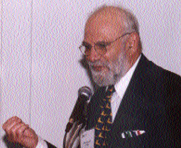
“There are other approaches to Parkinson’s disease complementary to these neurochemical approaches that are as important, as intriguing and maybe as complex,” said Oliver Sacks, who spoke after Greengard. Widely known for his depictions of patient stories in such books as Awakenings and The Man Who Mistook His Wife for a Hat, Sacks described how many Parkinson’s disease patients create their own means of handling the difficulties posed by their disease.
“Parkinson’s disease patients discover and utilize coping strategies that may not always be explainable in chemical terms,” he said. “I want to emphasize the importance of the individual’s experience, and his ability to articulate it.”
Many Parkinson’s patients find themselves aided by external stimuli such as music, for example, or by patterns and objects on the ground, Sacks said. Very early on, Charcot described how some Parkinson’s patients would carry around small balls of paper. When they became “stuck” in their movements, dropping one of the balls was all the impetus they needed to get going again.
“Parkinson’s patients have difficulty generating their own rhythm,” explained Sacks. “There seem to be fundamental disorders in the estimation of time and internal tempo.” That’s why external cues such as music and patterns, or a series of horizontal lines on the floor, can help such patients move more freely.
A Fascinating Neuroscientific Question
Over time, patients take the external stimuli that they have identified as helpful and internalize them, continued Sacks. They recall a visual image of the stimulus, rather than require the stimulus itself, and make it work for them. He recounted the story of a Parkinson’s disease patient who relied on a tree painted above his bed. Each morning he would visualize himself reaching for one of the branches, and he would be able to get out of bed. The mere image of grabbing the branch was sufficient to get him started each day.
So what is happening in the brain in these situations? “It is a neuroscientific question of a fascinating sort,” said Sacks. “It appears that the dopamine-depleted basal ganglia are somehow being bypassed” by these patients’ coping strategies. Indeed, modern neuroimaging techniques, such as positron emission tomography (PET) scanning and functional magnetic resonance imaging (fMRI), may someday yield the answer to this question.
Sacks concluded, “The power of the individual to articulate his experience, and explore it and analyze it, and the power of new forms of imaging and recording are going to transform neuroscience in the next 50 years.”
Also read: Music on the Mind: A Neurologist’s Take