In 2016, The New York Academy of Sciences and Takeda Pharmaceuticals announced the establishment of the Innovators in Science Award. The Award recognizes a promising Early-Career Scientist’s and an outstanding Senior Scientist’s contributions to biomedical science and is intended to support their commitment to innovative research. Two prizes of US$200,000 are awarded each Award cycle, in a specific therapeutic area, one to an Early-Career Scientist and the other to a well-established Senior Scientist from around the world. The winning scientists have distinguished themselves for the creativity and impact of their research. See https://nyaspubs.onlinelibrary.wiley.com/doi/toc/10.1111/(ISSN)1749-6632.innovators-science-award.
Early-career scientist, outstanding senior scientist each to receive US$200,000 in program sponsored by Takeda Pharmaceuticals
New York, NY | April 14, 2021 – The New York Academy of Sciences (NYAS) has opened nominations for the 2022 Innovators in Science Award, which will recognize significant achievement among early-career and senior scientists in the field of gastroenterology. This marks the first time scientists engaged in transformative research in gastroenterology will be eligible for the award, administered by the Academy and sponsored by Takeda Pharmaceuticals.
The program accepts nominations from eligible research institutions around the world to recognize the work of a promising early-career scientist and an outstanding senior scientist. Winners in each category will receive an unrestricted award of US$200,000 for having distinguished themselves for the creativity and impact of their research.
The Academy is accepting nominations through May 27, 2021, from more than 400 international universities and academic institutions, select government-affiliated and non-profit research institutions and the program’s Scientific Advisory Council, composed of renowned science and technology leaders. Candidates must be nominated by their institution and may not be self-nominated.
A judging panel composed of scientists, clinicians and international experts in gastroenterology will determine the two winners based on the quality, impact, novelty and promise of their research. They will be announced in January and honored at the 2022 Innovators in Science Award ceremony and symposium, scheduled for March 28-29, 2022, in Tokyo, Japan, as health and travel conditions allow.
“After one of the most challenging years of our time, recognizing and celebrating advancements in science is more important than ever,” said Nicholas B. Dirks, President and CEO of The New York Academy of Sciences. “The world is seeing firsthand how innovative science and thinking can improve human health, and we are committed to honoring those who are leading the way. The Innovators in Science Award salutes ground-breaking researchers who have developed science-based solutions to debilitating diseases, improving quality of life for people all over the world.”
Since its inception, the Innovators in Science Award has focused on acknowledging outstanding research and contributions in fields of medicine aligned with Takeda’s core therapeutic areas. The inaugural award recognized neuroscience discovery, followed the next year by regenerative medicine, rare disease research in 2020 and the latest on research in gastrointestinal and liver diseases. Recent research shows that 20-40% of adults worldwide are affected by at least one functional gastrointestinal disorder, which can dramatically impact quality of life.
Nominations may be submitted by representatives from the nominating institution through the Innovators in Science Award website via its online submission platform: https://innovatorsinscienceaward.smapply.io. Please refer to the guidelines and FAQ sections for other details on eligibility, nomination materials and the selection process.
By definition, a rare disease is one that afflicts relatively few people compared to the general population. Collectively, though, there are over 7,000 of these conditions known, causing immense suffering for an estimated 300 million patients. Because most rare diseases stem from specific genetic mutations, they’ve proven difficult to treat.
Genome sequencing and molecular medicine might soon change those grim statistics, though. For example, using short DNA or RNA sequences complementary to the messenger RNA for a gene, researchers can inhibit the expression of the associated protein. These complementary sequences—called antisense oligonucleotides—could soon be delivered as drugs to treat many rare diseases.
On October 2, 2020, The New York Academy of Sciences and Takeda Pharmaceuticals hosted the Frontiers in Rare Diseases: 2020 Innovators in Science Award Symposium, an event highlighting breakthroughs in rare diseases research and honoring 2020 Innovators in Science Award Winners Adrian Krainer, PhD and Jeong Ho Lee, MD, PhD. Presentations, a panel discussion, and a virtual poster session covered the basic science, recent clinical breakthroughs, and remaining challenges in this rapidly evolving field.
Symposium Highlights
While many rare diseases are inherited, others arise through mutations in somatic cells during life.
Antisense oligonucleotides can alter the expression of specific genes, potentially mitigating or reversing many genetic diseases.
Clinical trials for rare disease therapies must be tailored to the pathogenesis of each disease.
Redirecting neural stem cells to become neurons could treat many neurodegenerative diseases.
The COVID-19 pandemic is inspiring new collaborations that could be adapted to rare disease research.
Speakers
Jeong Ho Lee, MD, PhD Korea Advanced Institute of Science and Technology
Adrian Krainer, PhD Cold Spring Harbor Laboratory
Annemieke Aartsma-Rus, PhD Leiden University Medical Center
Don Cleveland, MD, PhD University of California, San Diego
Huda Zoghbi, MD Jan and Dan Duncan Neurological Research Institute, Texas Children’s Hospital
Brad Margus Cerevance
Graciana Diez-Roux, PhD Telethon Institute of Genetics and Medicine
David Fajgenbaum, MD University of Pennsylvania
Anne Heatherington, PhD Takeda Pharmaceuticals
Sponsors
The Winner’s Circle
Speakers
Jeong Ho Lee, MD, PhD Korea Advanced Institute of Science and Technology
Adrian Krainer, PhD Cold Spring Harbor Laboratory
Not Born This Way
Jeong Ho Lee, Early-Career Scientist winner of the 2020 Innovators in Science Award, discussed his work studying how somatic cell mutations—mutations that occur after development, during the normal process of cell division—result in rare neurological diseases caused by somatic cell mutations in the brain. Much of the recent boom in work on genetic diseases has focused on germline mutations. Because these mutations occur early in embryonic development, they show up in many types of cells throughout the body and are passed on to future offspring. These rare germline mutations can often be identified by sequencing the genomes of cells in easily accessible tissues, such as blood or skin. With advances in next-generation sequencing, “it’s become much easier to identify the germline mutations coding for many rare neurological disorders,” said Lee.
Somatic cell mutations occur throughout life, in every part of the body.
Nonetheless, germline mutations account for only a minority of rare neurological disorders. For example, 98% of epilepsy cases cannot be explained by germline mutations.
“We hypothesized that somatic cell mutations may be responsible for these unexplained neurological [diseases],” said Lee.
Somatic cell mutations occur during the ordinary cell division process that takes place billions of times in developing embryos, and continues to occur throughout life as somatic, or non-gamete, cells turn over.
DNA replication isn’t perfect, and human cells average 0.1 to 3 mutations per genome every time they divide. Lee theorized that a patient who hadn’t inherited an epilepsy-causing germline mutation might instead acquire somatic cell mutations in a subset of brain cells during development or later in life. If that happened, the mutation would only show up in the affected area of the brain, not in any other cells of the body.
One treatment for certain types of epilepsy is to resect the portion of the brain causing the seizures. Lee and his colleagues took samples of the brain tissue resected in these operations, along with blood samples from the same patients, and performed deep DNA sequencing to identify somatic mutations that occurred only in the affected brain tissue, not in the blood. They identified such mutations, including ones unique to genes involved in motor nerve activity, in 30% of the patients. When the scientists introduced the same mutations into a small percentage of neurons in developing mice, the animals developed epilepsy.
Next, the investigators looked at brain tumors, which can also cause epilepsy. One rare brain tumor type involves both glial cells and neurons, triggering epilepsy. Sequencing genetic information from cells in the tumors revealed that in 46% of affected patients, the glial cells and the neurons in the tumor shared an identical mutation.
“It means that the…neural stem cell already contained this… mutation,” said Lee, “and differentiated into the neuron and the glial cell.”
That could explain the high rate of disease recurrence in patients with these tumors; even if surgeons remove the entire tumor, the mutant stem cells might continue to produce more defective neurons and glial cells, which could then seed the growth of a new tumor.
To confirm that, Lee’s team collected an additional round of samples, this time sequencing cells not only from resected brain tumors and blood, but also from the subventricular zone in each patient’s brain, an area rich in undifferentiated neural stem cells. They found the tumor-associated mutation in the subventricular zone samples as well as the tumors, indicating that the error occurred in the neural stem cells, whose neuronal and glial descendants then migrated to where the tumor grew.
The researchers are also looking at neurodegenerative disorders such as Alzheimer’s disease.
“We hypothesized that somehow brain somatic mutation maybe accumulates over aging, and maybe associates with [Alzheimer’s disease development],” said Lee.
By performing deep sequencing on brain tissues from patients with and without Alzheimer’s disease, he and his colleagues identified somatic mutations unique to the diseased brains, supporting their theory.
In addition to identifying the underlying mechanisms behind neurological diseases, Lee is trying to help patients in other ways. In one effort, he has begun providing his results to clinicians to use in genetic counseling. Because conditions caused by somatic mutations aren’t heritable, while those caused by germline mutations are, patients who might be considering having children need to know which category they’re in.
The investigators are also trying to find ways to repair or mitigate the effects of somatic mutations in the brain, but it’s a tall order.
“Even if we found a molecular genetically validated target in the patient’s brain, it would be very difficult to develop a traditional drug to penetrate the blood-brain barrier and regulate the target,” Lee explained.
Instead, he’s hopeful that chemically modified strings of nucleic acids, called antisense oligonucleotides, will be able to target the somatic mutations he’s identified.
“I believe in the next five, ten, or twenty years, we probably can solve a lot of the rare neurological disorders,” he said.
Different Diseases, Different RNA Splices
Adrian Krainer won the 2020 Innovators in Science Senior Scientist Award, recognizing years of work spent developing treatments for rare diseases. Krainer and his colleagues were the first to develop an effective drug to treat spinal muscular atrophy. Affecting about 1 in 10,000 people worldwide, spinal muscular atrophy is an inherited genetic disease caused by a defect in the SMN1 gene. SMN1 encodes the SMN protein, which is essential for motor neuron survival. Patients with the mutation experience progressive loss of motor neurons, leading to loss of muscle control and, in most forms of the disease, early death.
Another gene, SMN2, also encodes the SMN protein, but cells usually splice out one of the protein coding sequences, or exons, from the SMN2 messenger RNA, preventing it from making the full-length protein. As a result, 80-90% of the protein translated from SMN2 RNA is truncated, nonfunctional, and rapidly degraded by the cell. Krainer reasoned that preventing the exon-skipping event might allow patients’ unmutated SMN2 genes to produce more functional SMN protein, overcoming the deficit caused by their mutated SMN1 genes. To do that, his team turned to antisense oligonucleotides, which encode the complementary, or antisense, sequence of a specific RNA target. When introduced into a cell, the antisense oligonucleotide binds specifically to its target sequence, triggering various cellular responses.
By designing an antisense oligonucleotide that altered the splicing of SMN2 messenger RNA, the researchers were able to get SMN1-mutant cells to produce more functional SMN protein. Subsequent preclinical and clinical trials proved that their antisense oligonucleotide also works in spinal muscular atrophy mouse models and in patients, respectively, significantly mitigating their motor neuron losses.
“Therefore this is a way that allows them to make closer to normal levels of a functional SMN protein in the presence of this drug,” said Krainer.
The oligonucleotide, now sold as nusinersen (Spinraza), was approved in the US in 2016 and the EU in 2017. Over 11,000 patients now receive it worldwide.
Nusinersen (Spinraza) pioneered many aspects of molecular medicine.
Based on the success of nusinersen, Krainer and his colleagues have begun looking at other RNA processing events to target with antisense oligonucleotides. One project focuses on familial dysautonomia, an inherited genetic disorder that affects only 310 known patients worldwide. These individuals have profound defects in their sensory neurons and autonomic nervous system, leading to symptoms that range from insensitivity to pain to difficulty swallowing.
“It is a very severe disease, a rare disease with a complex set of symptoms,” said Krainer, adding that “median survival is about 40 years of age.”
The condition is caused by a mutation in the gene for a protein called ELP1. As in SMN2, the mutation causes one exon of the gene’s messenger RNA to be spliced out, leading to a loss of functional ELP1 protein.
“So, we started targeting this aberrant splicing event using the same screening strategy and the same chemistry that we used…for spinal muscular atrophy,” said Krainer.
That effort identified an antisense oligonucleotide that can reverse the ELP1 RNA splicing defect in cultured cells from patients, as well as a transgenic mouse model.
“We feel that this is ready for clinical development; it is a challenge, though, because of the rarity of this disease,” said Krainer.
With only a few hundred patients in the US and Israel, the market for familial dysautonomia therapies is minuscule, and effective screening of potential carriers of the affected gene has led to very few new patients being born.
Not all RNA splicing-related diseases are rare, though. Work by several researchers has shown that in at least some cases, a change in the splicing of messenger RNA can help cancer cells grow. Alternatively, spliced forms of the messenger RNA for the PKM gene can produce two different isoforms of the metabolic enzyme pyruvate kinase. PKM1 predominates in normal adult tissues, while tumors and some developing tissues favor PKM2 production.
Using the same approach that worked in their rare disease work, Krainer’s team screened antisense oligonucleotides and identified candidates that bound the PKM messenger RNA and directed its splicing to favor PKM1 protein production. Putting these oligonucleotides into hepatocellular carcinoma cells causes the cells to shift their metabolism and slow their growth. In a mouse model of hepatocellular carcinoma, injecting the antisense oligonucleotides led to a significant reduction in tumor growth compared to control animals treated with saline solution.
Annemieke Aartsma-Rus, PhD Leiden University Medical Center
Don Cleveland, MD, PhD University of California, San Diego
The Kindest Cut
Annemieke Aartsma-Rus began the meeting’s third session with a presentation about her group’s efforts to address Duchenne muscular dystrophy with antisense oligonucleotides. While Krainer’s approach to rare diseases focuses on conditions where an exon needs to be added back into a messenger RNA, Aartsma-Rus described a case where it’s better to remove one.
Duchenne muscular dystrophy is an X-linked genetic disorder. In most cases, a mutation in the dystrophin gene shifts the messenger RNA’s reading frame, causing translation of the dystrophin protein to fail.
“Patients become wheelchair dependent around the age of 12, need assisted ventilation around the age of 20, and generally die in the second to fourth decade of life,” said Aartsma-Rus.
A related but milder disorder, Becker muscular dystrophy, also involves a deletion in the dystrophin gene but doesn’t shift the messenger RNA’s reading frame. As a result, patients with Becker muscular dystrophy produce partially functional dystrophin and exhibit a slower disease progression.
Skipping an exon in the RNA can fix a frame-shift mutation.
Looking at the affected DNA and RNA sequences, Aartsma-Rus reasoned that most Duchenne muscular dystrophy patients could make Becker-like dystrophin, if their cells could simply skip the affected exon in their dystrophin messenger RNA. To test that, she and her colleagues developed chemically modified antisense oligonucleotides that would remain stable in blood and tissues, and began testing them as potential drugs. By designing an oligonucleotide that targeted RNA splicing, the team restored dystrophin expression in cultured cells carrying a Duchenne muscular dystrophy mutation.
The researchers discovered a potential roadblock in a mouse model: antisense oligonucleotides injected into the animals’ tail veins were absorbed almost entirely by the liver and kidneys. The investigators could inject the molecules directly into muscles instead, but that clearly wouldn’t be a practical way to deliver treatment to patients.
“We have over 700 different muscles, and you’ll have to treat patients repeatedly,” said Aartsma-Rus, “so local injection of each and every muscle weekly or even monthly is likely not realistic.”
However, in a mouse model of Duchenne muscular dystrophy, the team discovered that oligonucleotides injected into the animals’ tail veins were absorbed into muscles ten times better than they had been in wild-type mice.
“The first time we thought we’d made a mistake, so we repeated it a couple of times,” said Aartsma-Rus, “but every time we saw that there was higher uptake by the dystrophic muscle than the healthy muscle.”
Dystrophin deficiency causes muscle cells to become more permeable, leading to leakage of cellular components, but this leakage works in both directions; the dystrophic cells readily absorbed oligonucleotides that healthy cells excluded.
Flush with this preclinical victory, the team began setting up clinical trials in 2007. The initial multi-center, open-label trial found that the antisense oligonucleotides caused no serious side effects, and eight of the twelve patients tested saw their conditions remain stable throughout the trial. To evaluate efficacy, the investigators moved into a phase 2b trial, which continued to show dose-dependent effectiveness in treated patients. However, a larger phase 3 trial yielded disappointment, with no significant difference in outcomes between treated and control patients.
“So, what happened [to explain why] we see these beneficial effects in the phase 2 trial, but in the phase 3 trial we see no effect?” Aartsma-Rus asked.
Analyzing the results and the disease further, she realized that the trials were built on the flawed assumption that the patients’ progression would be linear. Instead, they realized that younger patients tend to remain stable for an extended period, followed by a rapid decline later in life. By mixing different ages in the phase 3 trial, patients with worse disease symptoms likely masked any treatment benefits in those with milder symptoms.
Looking at the trial’s failure, Aartsma-Rus concluded that she and her colleagues should have opened discussions with regulatory agencies sooner, and studied the natural history of the disease more thoroughly, before initiating the phase 3 study. Unfortunately, the expensive late-stage failure has soured companies on further clinical development of exon-skipping antisense oligonucleotides for Duchenne muscular dystrophy. Aartsma-Rus has since focused on preventing such an outcome in the future.
“Now we have an open dialogue with academics, with patients, with regulators in the EU, and it is also starting in the US, developing new outcome measures,” she said, adding that “future trials will be better.”
Batting for Lou Gehrig
Don Cleveland discussed his group’s efforts to treat neurodegenerative diseases in the brain, especially those that develop gradually with age. In many of these conditions, such as Alzheimer’s and Parkinson’s disease and amyotrophic lateral sclerosis, “the genes that contribute to disease are all widely expressed…throughout the nervous system, not within individual classes of neurons,” said Cleveland. Mechanistic studies have suggested that decreasing the expression of the defective gene products in some of these cells could moderate the course of disease, so Cleveland and his colleagues set out to do just that.
The researchers first focused on amyotrophic lateral sclerosis (ALS), also known as Lou Gehrig’s disease. About four to five million people alive today will die of ALS, a progressive neurodegenerative condition that can be inherited or occur spontaneously in adults. One inherited form of the disease stems from a mutation in the gene for superoxide dismutase, which causes neurons to die through mechanisms that aren’t entirely clear yet.
Using the same strategy as his co-speakers, Cleveland’s lab designed antisense oligonucleotides that bind specifically to the superoxide dismutase messenger RNA and target it for degradation in the cell. That decreases the level of the enzyme, an intervention that had previously been shown to ameliorate ALS progression in a mouse model of the disease. The next challenge was delivering the oligonucleotides to affected neurons in the brain.
Antisense oligonucleotides have immense potential to be used as drugs against a wide range of diseases.
“These DNA drugs were ten to fifteen times the size of a typical drug, and they’re heavily charged,” said Cleveland, “so the pharmacology textbooks all said that there was no uptake mechanism that would permit them to be efficiently taken up [by neurons].” Nonetheless, he continued, “we tried it anyway, and it turns out that the cells of the nervous system hadn’t read the textbooks.”
Injecting the antisense oligonucleotides into the cerebrospinal fluid of mice genetically modified to develop severe ALS doubled the animals’ survival times.
While the superoxide dismutase defect was the first ALS mutation discovered, the most common cause of the inherited form of the disease is a mutation in a gene called ORF72, which inserts extra nucleotides into a non-coding region of the gene. This causes defective messenger RNA to accumulate, killing neurons because of a lack of functional ORF72 gene products and the accumulation of toxic byproducts of the altered gene. Antisense oligonucleotides targeting the defective RNA, however, inhibit its accumulation without reducing the production of working ORF72 gene products in cultured cells.
In an animal model of the ORF72 defect, the results were even more impressive.
“We dosed these animals [with the antisense oligonucleotides] at the age of disease onset and asked what happens, and the answer is we prevented further disease development for the life of those animals with a single dose injection applied at the initial signs of disease,” said Cleveland.
His team initiated clinical trials on this therapy in 2018, just seven years from the date when researchers had first published the data showing the ORF72 mutation caused ALS.
Although it’s an important target for research, inherited ALS accounts for only 10% of the disease’s total cases. In 90% of patients, the condition develops spontaneously due to somatic cell mutations later in life. Many of these cases involve mutations in the TDP-43 gene, which encodes a nuclear protein that regulates Stathmin-2, which in turn plays a critical role in regulating the cytoskeleton in neurons. TDP-43 normally binds the Stathmin-2 precursor RNA and ensures that it gets spliced properly into messenger RNA. Mutations that inactivate TDP-43 cause a loss of functional Stathmin-2, which is a hallmark of sporadic ALS.
Using cultured neurons, Cleveland and his colleagues found that a properly designed antisense oligonucleotide could compensate for the loss of TDP-43 activity, restoring normal RNA splicing and Stathmin-2 expression.
“This now enables a strategy for therapy for sporadic ALS,” said Cleveland.
If the result holds in other preclinical models, he expects to take that approach into clinical trials in 2023.
Besides correcting specific defects within a cell, antisense nucleotides can potentially redirect a cell’s fate entirely. That’s the central theme of another project in Cleveland’s lab, in which the team is causing astrocytes to change their identities. Astrocytes are companion cells in the nervous system that arise from the same stem cells as neurons. Using antisense oligonucleotides, the investigators can suppress two genes that direct cells into the astrocyte lineage, causing them to become neurons instead. Cleveland is initially focusing on treating Parkinson’s disease with this approach, but he explained that “this…conversion of astrocytes into replacement neurons may be broadly applicable for neurogenic disease.”
Huda Zoghbi Jan and Dan Duncan Neurological Research Institute, Texas Children’s Hospital
Maybe Not So Rare
Huda Zoghbi gave the meeting’s keynote presentation, which covered her work on Rett syndrome. Caused by spontaneous mutations in the MECP2 gene on the X chromosome, Rett syndrome is a progressive neurodegenerative disease that primarily manifests itself in girls. MECP2 is critical for gene regulation in neurons. Because females carry two copies of the X chromosome and an inactivate one in each cell, an inactivating mutation in MECP2 impairs the function of 50% of the affected individual’s neurons. That manifests itself as a rapid regression in motor and cognitive abilities by age two.
In boys, who have only one X chromosome, inactivation of MECP2 is generally lethal before age two. They don’t live long enough to develop the classic symptoms of Rett syndrome. However, recent work has revealed that some males acquire mutations that cause less severe defects in MECP2.
“What we’ve learned is when people carry milder mutations, we will see milder phenotypes, such as mild learning disability with…neuropsychiatric features,” said Zoghbi.
These individuals’ phenotypes can range from autism to hyperactivity or schizophrenia, but most die by middle age due to neurodegeneration. Females with mild defects in MECP2 show non-random inactivation of their X chromosomes, favoring the healthy copy of the gene and enabling them to develop and live normally. Some patients also have duplications in their MECP2 genes, often leading to severe neurological problems and premature death.
To understand the mechanisms driving Rett syndrome, Zoghbi and her colleagues developed a series of genetically modified mice carrying various duplications or mutations in MECP2. Consistent with the findings in humans, these animals display a spectrum of phenotypes depending on the severity of their MECP2 disruptions.
“The brain is very sensitive to the activities and functions of this protein, and we’ve done a lot of studies on both the loss and the gain models,” said Zoghbi.
She and her colleagues found that all types of neurons require functional MECP2 to operate normally.
Mutations affecting different types of neurons can cause a wide range of neurological phenotypes.
Next, Zoghbi and her colleagues tried inactivating MECP2 in excitatory and inhibitory neurons separately. They found that in both cases, animals developed obesity, lost motor coordination, and died young. However, targeting MECP2 only in inhibitory neurons led to more learning and social defects in the animals, while inactivating it only in excitatory neurons caused more anxiety and tremors. These phenotypes represent the downstream effects of the genes MECP2 would normally regulate.
“Given that it’s important for practically every cell, really there’s two major ways you can think of treating this disorder, either gene replacement therapy…or perhaps exploring modulation of the [MECP2 regulatory] circuit,” said Zoghbi.
Taking the latter approach, the investigators implanted electrodes into the brains of mice to deliver small electrical pulses. This type of deep brain stimulation, which has been shown to reverse many types of neuronal signaling and development defects, is already approved for human treatment of several neurological disorders. Stimulating the brains of Rett syndrome model mice leads to significant recovery in their learning, memory, and motor abilities that persists for weeks after treatment.
“It was really quite a dramatic rescue in that all these phenotypes normalize, and their normalization…lasted for several weeks,” said Zoghbi.
The treated animals’ neurons also displayed gene expression patterns similar to wild-type animals, whereas untreated animals showed significant gene dysregulation.
“The Rett brain, at least in mice, is responsive to neuromodulation,” said Zoghbi.
Looking at the MECP2 gene itself, Zoghbi’s team identified regulatory sequences that control its expression level. Altering these sequences to increase or decrease the amount of MECP2 expressed in mice underscored their earlier findings, showing that even modest changes in MECP2 levels led to detectable neurological phenotypes.
Like other speakers at the meeting, Zoghbi and her colleagues are also exploring the potential of antisense oligonucleotides as therapies. That approach seems especially promising for patients with duplications in MECP2 that lead to overexpression of the gene. In mice that recapitulate this condition, the researchers found that treatment with antisense oligonucleotides against MECP2 could reduce the amount of functional protein in neurons down to wild-type levels. The treatment reversed the animals’ motor defects.
Titrating the antisense oligonucleotide dosage also revealed that even modest decreases in excess MECP2 can lead to major improvements in symptoms.
“If you can even partially decrease the protein…you will probably rescue quite a bit of the features of the disease,” Zoghbi said. She added, “I’ve really never worked with a protein that is so exquisitely sensitive to the levels.”
Graciana Diez-Roux, PhD Telethon Institute of Genetics and Medicine
David Fajgenbaum, MD University of Pennsylvania
Anne Heatherington, PhD Takeda Pharmaceuticals
Silver Linings
The meeting’s general session concluded with a panel discussion led by Brad Margus, co-founder and CEO of Cerevance. With a background in business, Margus moved into rare disease drug development after his daughter was diagnosed with ataxia-telangiectasia, a genetic disorder that causes neurodegeneration and immune dysfunction. The panel also featured Graciana Diez-Roux, chief scientific officer at the rare disease-focused Telethon Institute; David Fajgenbaum, a physician-scientist who both studies and suffers from Castleman syndrome; and Anne Heatherington, a data scientist for Takeda Pharmaceuticals with extensive experience studying Duchenne muscular dystrophy.
Panel members discussed the need for improvement in collaborations between patients and researchers.
“There is a lot of miscommunication within the rare disease research space, [but] I think there’s been a really great trend for groups like Takeda and others toward engaging patients in the research process,” said Fajgenbaum, adding that “I also think clinicians can really be a part of this.”
Besides improving clinical trial recruitment, involving patients more directly in research can have far-reaching benefits for scientists.
“It’s incredible how our PhD students, when they have the chance [to interact] with the patients and [get] to know the patients’ organizations…how their motivation and their love for what they do changes,” Diez-Roux said.
Besides increasing collaborations between patients and scientists, all of the panelists endorsed the need for strong, well-defined partnerships with pharmaceutical companies. Margus described his company’s efforts to improve data collection and sharing for ataxia-telangiectasia, which included building a system that uses wearable devices to collect movement data from patients around the clock.
“The data [are] truly owned by the families and the community, and we can make decisions about sharing the data with academics or any researcher in the world in a matter of days,” said Margus.
Good partnerships require more than just good databases, though. Academic researchers accustomed to independent, curiosity-driven experimental design and flexible deadlines sometimes have trouble accommodating pharmaceutical companies’ urgent, goal-directed needs.
“I think the model has to be somewhere between…the industry knowing how to deal with the academic research and academic researchers being open to notice that industries have…different goals in some respects,” said Diez-Roux.
The group also discussed the impact of the COVID-19 pandemic. In the short term, of course, the global shutdown caused by the SARS-CoV-2 virus has halted or delayed many rare disease studies. However, panelists agreed that some of the innovative approaches developed for the pandemic response could transform many aspects of rare disease research in the future.
“We have been very involved in a lot of the COVID alliances, and have been steeped in novel ways of working,” said Takeda’s Heatherington.
As an example, she pointed to the company’s involvement in multi-corporation consortia to develop new therapies and even entirely new platforms for therapies.
“That’s a real breakthrough in terms of how we do our business, that extent of collaboration for [competitors to] come together,” Heatherington continued.
At the same time, “the public is realizing more how important research is, and this goes for COVID, but I think it goes for all diseases,” said Diez-Roux. Both she and Heatherington also pointed out that the pandemic has underscored the potential tradeoffs between speed and safety in therapeutic development, and highlighted the importance of oversight in clinical trials.
The meeting concluded with a virtual poster session, featuring rapid-fire presentations of some of the newest research in rare diseases and offering attendees the ability to interact with the presenters directly. Like the other presentations, the posters represented the diversity and enthusiasm of rare disease researchers.
“What makes me optimistic is the passion and the knowledge…and the fact that we have people that are so dedicated to rare diseases,” said Heatherington.
The 2020 Innovators in Science Award winners include a biochemist/molecular geneticist from Cold Spring Harbor Laboratory and brain disorder researcher from the Korea Advance Insitute of Science and Technology.
New York, NY | July 8, 2020 and Osaka, Japan | July 8, 2020 – Takeda Pharmaceutical Company Limited (“Takeda”) (TSE:4502) and the New York Academy of Sciences announced today the Winners of the third annual Innovators in Science Award for their excellence in and commitment to innovative science that has significantly advanced the field of rare disease research. Each Winner receives a prize of US $200,000.
Senior Scientist Award: Adrian R. Krainer
The 2020 Winner of the Senior Scientist Award is Adrian R. Krainer, Ph.D., St. Giles Foundation Professor at Cold Spring Harbor Laboratory. Prof. Krainer is recognized for his outstanding research on the mechanisms and control of RNA splicing, a step in the normal process by which genetic information in DNA is converted into proteins. Prof. Krainer studies splicing defects in patients with spinal muscular atrophy (SMA), a devastating, inherited pediatric neuromuscular disorder caused by loss of motor neurons, resulting in progressive muscle atrophy and eventually, death. Prof. Krainer’s work culminated notably in the development of the first drug to be approved by global regulatory bodies that can delay and even prevent the onset of an inherited neurodegenerative disorder.
“Collectively, rare diseases affect millions of families worldwide, who urgently need and deserve our help. I’m extremely honored to receive this recognition for research that my lab and our collaborators carried out to develop the first approved medicine for SMA,” said Prof. Krainer. “As basic researchers, we are driven by curiosity and get to experience the thrill of discovery; but when the fruits of our research can actually improve patients’ lives, everything else pales in comparison.”
Early-Career Scientist Award: Jeong Ho Lee
The 2020 Winner of the Early-Career Scientist Award is Jeong Ho Lee, M.D., Ph.D, Associate Professor, Korea Advanced Institute of Science and Technology (KAIST). Prof. Lee is recognized for his research investigating genetic mutations in stem cells in the brain that result in rare developmental brain disorders.
He was the first to identify the causes of intractable epilepsies and has identified the genes responsible for several developmental brain disorders, including focal cortical dysplasias, Joubert syndrome—a disorder characterized by an underdevelopment of the brainstem—and hemimegalencephaly, which is the abnormal enlargement of one side of the brain. Prof. Lee also is the Director of the National Creative Research Initiative Center for Brain Somatic Mutations, and Co-founder and Chief Technology Officer of SoVarGen, a biopharmaceutical company aiming to discover novel therapeutics and diagnosis for intractable central nervous system (CNS) diseases caused by low-level somatic mutation.
“It is a great honor to be recognized by a jury of such globally respected scientists whom I greatly admire,” said Prof. Lee. “More importantly, this award validates research into brain somatic mutations as an important area of exploration to help patients suffering from devastating and untreatable neurological disorders.”
The 2020 Innovators in Science Award Ceremony and Symposium
The 2020 Winners will be honored at the virtual Innovators in Science Award Ceremony and Symposium in October 2020. This event provides an opportunity to engage with leading researchers, clinicians and prominent industry stakeholders from around the world about the latest breakthroughs in the scientific understanding and clinical treatment of genetic, nervous system, metabolic, autoimmune and cardiovascular rare diseases.
“At Takeda, patients are our North Star and those with rare diseases are often underserved when it comes to the discovery and development of transformative medicines,” said Andrew Plump, M.D., Ph.D., President, Research & Development at Takeda. “Insights from the ground-breaking research of scientists like Prof. Krainer and Prof. Lee can lead to pioneering approaches and the development of novel medicines that have the potential to change patients’ lives. That’s why we are proud to join with the New York Academy of Sciences to broadly share and champion their work — and hopefully propel this promising science forward.”
“Connecting science with the world to help address some of society’s most pressing challenges is central to our mission,” said Nicholas Dirks, Ph.D., President and CEO, the New York Academy of Sciences. “In this third year of the Innovators in Science Award we are privileged to recognize two scientific leaders working to unlock the power of the genome to bring innovations that address the urgent needs of patients worldwide affected by rare diseases.”
About the Innovators in Science Award
The Innovators in Science Award grants two prizes of US $200,000 each year: one to an Early-Career Scientist and the other to a well-established Senior Scientist who have distinguished themselves for the creative thinking and impact of their research. The Innovators in Science Award is a limited submission competition in which research universities, academic institutions, government or non-profit institutions, or equivalent from around the globe with a well-established record of scientific excellence are invited to nominate their most promising Early-Career Scientists and their most outstanding Senior Scientists working in one of four selected therapeutic fields of neuroscience, gastroenterology, oncology, and regenerative medicine.
Prize Winners are determined by a panel of judges, independently selected by The New York Academy of Sciences, with expertise in these disciplines. The New York Academy of Sciences administers the Award in partnership with Takeda.
For more information please visit the Innovators in Science Award website.
About Takeda Pharmaceutical Company Limited
Takeda Pharmaceutical Company Limited (TSE:4502/NYSE:TAK) is a global, values-based, R&D-driven biopharmaceutical leader headquartered in Japan, committed to bringing Better Health and a Brighter Future to patients by translating science into highly-innovative medicines. Takeda focuses its R&D efforts on four therapeutic areas: Oncology, Rare Diseases, Neuroscience, and Gastroenterology (GI).
We also make targeted R&D investments in Plasma-Derived Therapies and Vaccines. We are focusing on developing highly innovative medicines that contribute to making a difference in people’s lives by advancing the frontier of new treatment options and leveraging our enhanced collaborative R&D engine and capabilities to create a robust, modality-diverse pipeline. Our employees are committed to improving quality of life for patients and to working with our partners in health care in approximately 80 countries. For more information, visit https://www.takeda.com.
The human body regenerates itself constantly, replacing old, worn-out cells with a continuous supply of new ones in almost all tissues. The secret to this perpetual renewal is a small but persistent supply of stem cells, which multiply to replace themselves and also generate progeny that can differentiate into more specialized cell types. For decades, scientists have tried to isolate and modify stem cells to treat disease, but in recent years the field has accelerated dramatically.
A major breakthrough came in the early 21st century, when researchers in Japan figured out how to reverse the differentiation process, allowing them to derive induced pluripotent stem (iPS) cells from fully differentiated cells. Since then, iPS cells have become a cornerstone of regenerative medicine. Researchers can isolate cells from a patient, produce iPS cells, genetically modify them to repair any defects, then induce the cells to form the tissue the patient needs regenerated.
On April 26, 2019, the New York Academy of Sciences and Takeda Pharmaceuticals hosted the Frontiers in Regenerative Medicine Symposium to celebrate 2019 Innovators in Science Award winners and highlight the work of researchers pioneering techniques in regenerative medicine. Presentations and an interactive panel session covered exciting basic research findings and impressive clinical successes, revealing the immense potential of this rapidly developing field.
Symposium Highlights
New cell lines should reduce the time and cost of developing stem cell-derived therapies.
The body’s microbiome primes stem cells to respond to infections.
iPS cell-derived therapies have already treated a deadly genetic skin disease and age-related macular degeneration.
Polyvinyl alcohol is a superior substitute for albumin in stem cell culture media.
A newly isolated type of stem cell reveals the stepwise process driving early embryo organization.
Speakers
Shinya Yamanaka Kyoto University
Shruti Naik New York University
Michele De Luca University of Modena and Reggio Emilia
Masayo Takahashi RIKEN Center for Biosystems Dynamics Research
Hiromitsu Nakauchi Stanford University and University of Tokyo
Brigid L.M. Hogan Duke University School of Medicine
Emmanuelle Passegué Columbia University Irving Medical Center
Hans Schöler Max Planck Institute for Molecular Biomedicine
Austin Smith University of Cambridge
Moderator: Azim Surani University of Cambridge
Sponsors
Recent Progress in iPS Cell Research Application
Speakers
Shinya Yamanaka Kyoto University
Highlights
Current protocols for using induced pluripotent stem (iPS) cells clinically are slow and expensive.
HLA “superdonor” iPS cell lines can be used to treat multiple patients, reducing costs.
A unique academic-industry partnership is helping iPS cell therapies reach the clinic.
Faster, Cheaper, Better
Shinya Yamanaka of Kyoto University, gave the meeting’s keynote presentation, summarizing his laboratory’s recent work using induced pluripotent stem (iPS) cells for regenerative medicine. The first clinical trial using iPS cells to treat age-related macular degeneration started five years ago. In his clinical trial, physicians isolated somatic cells from a patient, then used developed culture techniques to derive iPS cells from them. iPS cells can proliferate and differentiate into any type of cell in the body, which can then be transplanted back into the patient. Experiments over the past five years have shown that this approach has the potential to treat diseases ranging from age-related macular degeneration to Parkinson’s disease.
However, this autologous transplantation strategy is slow and expensive. “It takes up to a year just evaluating one patient, [and] it costs us almost one million US dollars,” said Yamanaka. Before transplanting the differentiated cells, the researchers evaluated the entire iPS cell derivation and iPS cell differentiation processes, adding to time and cost. As another strategy, Yamanaka’s team is working on the iPS Cell Stock for Regenerative Medicine. Here, iPS cells are derived from blood cells of healthy donors, not the patients, and are stocked. The primary problem with this approach is the human leukocyte antigen (HLA) system, which encodes multiple cell surface proteins. Each person has a specific combination of HLA genes, or haplotype, defining the HLA proteins expressed on their own cells. The immune system recognizes and eliminates any cell expressing non-self HLA proteins. Because there are millions of potential HLA haplotypes, cells derived from one person will likely be rejected by another.
The homozygous “superdonor” cell line has limited immunological diversity, allowing it to match multiple patients.
The homozygous “superdonor” cell line has limited immunological diversity, allowing it to match multiple patients.
To address that, Yamanaka and his colleagues are collaborating with the Japanese Red Cross to develop “superdonor” iPS cells. These cells carry homozygous alleles for different human lymphocyte antigen (HLA) genes, limiting their immunological diversity and making them match multiple patients. So far, the team has created four “superdonor” cell lines, allowing them to generate cells compatible with 40% of the Japanese population. Those cells are now being used in clinical trials treating macular degeneration and Parkinson’s disease, with more indications planned.
“So far so good,” said Yamanaka, but he added that “in order to cover 90% of the Japanese population we would need 150 iPS cell lines, and in order to cover the entire world we would need over 1,000 lines.” It took the group about five years to generate the first four lines, so simply repeating the process that many more times isn’t practical.
Instead, Yamanaka hopes to take the HLA reduction a step further, knocking out most of the major HLA genes to generate cells that will survive in large swaths of the population. However, simply knocking out entire families of genes isn’t enough. Natural killer cells attack cells that are missing particular cell surface antigens, so the researchers had to leave specific markers in the cells, or reintroduce them transgenically. Natural killer and T cells from various donors ignore leukocytes derived from these highly engineered iPS cells, proving that the concept works. With this approach, just ten lines of iPS cells should yield a range of donor cells suitable for any human HLA combination. Yamanaka expects these gene-edited iPS cells to be available in 2020.
By 2025, Yamanaka hopes to announce “my iPS cell” technology. This technology will reduce the cost and time for autologous transplantation to about $10,000 and one month per patient.
While preclinical and early clinical trials on iPS cells have yielded promising results, the new therapies must still cross the “valley of death,” the pharmaceutical industry’s term for the unsuccessful transition and industrialization of innovative ideas identified in academia to routine clinical use. In an effort to make that process more reliable, Yamanaka and his colleagues have begun a unique collaboration with Takeda Pharmaceutical Company Limited, Japan’s largest drug maker. The effort involves 100 scientists, 50 each from the company and academic laboratories. The corporate researchers gain access to the latest basic science developments on iPS cell technology, while the academics can use the company’s cutting-edge R&D know-how equipment and vast chemical libraries.
In one project, the collaborators used iPS cells to derive pancreatic islet cells, and then encapsulated the cells in an implantable device to treat type 1 diabetes. The system successfully decreased blood glucose in a mouse model, and the team is now scaling up cell production to test it in humans in the future. Another effort identified chemicals in Takeda’s compound library that speed cardiomyocyte maturation, which the researchers are now using to improve iPS cell-derived treatments for heart failure. In a third project, the team has modified iPS cell-derived T cells to identify and attack tumors, again showing promising results in a mouse model.
Inflammation causes persistent changes in epithelial stem cells, priming them for subsequent immune responses.
Modified iPS cells can be used to cure a patient with a deadly genetic skin defect.
A small population of self-renewing stem cells maintains human skin cells.
Sparring Partners
Shruti Naik, Early-Career Scientist winner of the 2019 Innovators in Science Award, discussed her work on epithelial barriers. These barriers, which include skin and the linings of the gut, lungs, and urogenital tract, exhibit nuanced responses to the many microbes they encounter. Injuries and pathogenic infections trigger prompt inflammatory responses, but the millions of harmless commensal bacteria that live on these surfaces don’t. How does the epithelium know the difference?
To ask that question, Naik first studied germ-free mice, which lack all types of bacteria. These animals have defective immune responses against pathogens that affect epithelia, so commensal bacteria are clearly required for developing normal epithelial immunity. Naik inoculated the germ-free mice with bacterial strains found either on the skin or in the guts of normal mice, then assessed their immune responses in those two compartments.
“When you gave gut-tropic bacteria, you were essentially able to rescue immunity in the gut but not the skin, and conversely when you gave skin-tropic bacteria, you were able to rescue immunity in the skin and not the gut,” said Naik. Even though the commensal bacteria caused no inflammation, they did activate certain T cells in the epithelia they colonized, apparently preparing those tissues for subsequent attacks by pathogens.
Next, Naik took germ-free mice inoculated with Staphylococcus epidermidis, a normal skin commensal bacterium, and challenged them with an infection by Candida albicans, a pathogenic yeast. The bacterially primed mice produced a much more robust immune response against the yeast infection than control animals that hadn’t gotten S. epidermidis. Naik confirmed that this immune training effect operates through the T cell response she’d seen before. “You essentially develop an immune arsenal to your commensals that helps protect against pathogens,” Naik explained, adding that each epithelial barrier requires its own commensal bacteria to trigger this response.
Augmented wound repair in post-inflammation skin reveals that naive and inflammation-educated skin stem cells respond differently to subsequent stresses.
Augmented wound repair in post-inflammation skin reveals that naive and inflammation-educated skin stem cells respond differently to subsequent stresses.
The response to epithelial commensals is remarkably durable; Naik found that the skin T cells in the inoculated mice remained on alert a year after their initial activation. That led her to wonder whether non-hematopoietic cells, especially epithelial stem cells, contribute to immunological memory in the skin.
To probe that, Naik and a colleague used a mouse model in which the topical drug imiquimod induces a temporary psoriasis-like skin inflammation. By tracing the lineages of cells in the animals’ skin, the researchers found that epithelial stem cells expand during this inflammation, and then persist. Challenging the mice with a wound one month after the inflammation resolves leads to faster healing than if the mice hadn’t had the inflammation. Several other models of wound healing yielded the same result. The investigators concluded that naive and inflammation-educated skin stem cells respond differently to subsequent stresses.
Naik’s team found that inflammation causes persistent changes in skin stem cells’ chromatin organization. Using a clever reporter gene assay, they demonstrated that the initial inflammation leaves inflammatory gene loci more open in the chromatin, making them easier to activate after subsequent insults. “What was really surprising to us was that this change never fully resolved,” said Naik. Even six months after the acute inflammation, skin stem cells retained the distinct post-inflammatory chromatin structure and the ability to heal wounds quickly. This chronic ready-for-action state isn’t always beneficial, though. Naik noticed that the mice that had had the inflammatory treatment were more prone to developing tumors, for example.
In establishing her new laboratory, Naik has now turned her focus to another aspect of epithelial immunity: the link between immune responses and tissue regeneration. She looked first at a type of T cells found in abundance around hair follicles on skin. Mice lacking these cells exhibit severe delays in wound healing, apparently as a result of failing to vascularize the wound area. That implies a previously unknown role for inflammatory T cells in vascularization, which Naik and her lab are now probing.
Skin Deep
Michele De Luca, Senior Scientist winner of the 2019 Innovators in Science Award, has developed techniques for regenerating human skin from transgenic epidermal stem cells. Researchers first isolated holoclones, or cells derived from a single epidermal stem cell, over 30 years ago. These cells can be used to grow sheets of skin in culture for both research and clinical use, but scientists have only recently begun to elucidate how the process works.
The first stem cell-derived therapies tested in humans were for skin and eye burns, allowing doctors to regenerate and replace burned epidermal tissue from a patient’s own stem cells. That’s the basis of Holoclar, a stem cell-based treatment for severe eye burns approved in Europe in 2015.
Holoclar and similar procedures work well for injured patients with normal epithelia. “We wanted to genetically modify those cells in order to address one of the most important genetic diseases in the dermatology field, which is epidermolysis bullosa (EB), a devastating skin disease,” said De Luca. In EB, patients carry a genetic defect in cell adhesion that causes severe blisters all over their skin and prevents normal healing. A large number of EB patients die as children from the resulting infections, and those who survive seldom get beyond young adulthood before succumbing to squamous cell carcinomas.
De Luca developed a strategy to isolate stem cells from a skin biopsy, repair the genetic defect in these cells with a retroviral vector, and then grow new skin in culture that can be transplanted back to the patient, replacing their original skin with genetically repaired skin. In 2015, the researchers carried out the procedure on a young boy named Hassan, who had arrived in the burn unit of a German hospital with EB after fleeing Syria. The burn unit was only able to offer palliative care, and his prognosis was poor because of his constant blistering and infections. De Luca’s team received approval to perform their gene therapy on him.
The new strategy, which combines cell and gene therapy, resulted in the restoration of normal skin adhesion in Hassan.
After isolating and modifying epidermal stem cells from Hassan and growing new sheets of skin in culture, De Luca’s team re-skinned the patient’s arms and legs, then his abdomen and back. The complete procedure took about three months. The new skin resists blister formation even when rubbed and heals normally from minor wounds. In the ensuing three and a half years, Hassan has begun growing normally and living an ordinary, healthy life.
Detailed analysis of skin biopsies showed that Hassan’s epidermis has normal cellular adhesion machinery and revealed that his skin is now derived from a population of proliferating transgenic stem cells, with no single clone dominating. By tracing the lineages of cells carrying the introduced transgene, De Luca was able to identify self-renewing transgenic stem cells, intermediate progenitor cells, and fully differentiated stem cells, indicating normal skin growth and replacement.
Besides being good news for the patient, the results confirmed a longstanding theory of skin regeneration. “These data formally prove that the human epidermis is sustained only by a small population of long-lived stem cells that generates [short-lived epithelial] progenitors,” said De Luca, adding that “with this in mind, we’ve started doing other clinical trials.”
The researchers plan to continue targeting junctional as well as dystrophic forms of EB, both of which are genetically distinct from EB simplex. Initial experiments revealed that in these conditions, transplant recipients developed mosaic skin, where some areas continued to be produced from cells lacking the introduced genetic repair. The non-transgenic cells appeared to be out-competing the transgenic cells and supplanting them, undermining the treatment. De Luca and his colleagues developed a modified strategy that gave the transgenic cells a competitive advantage. This approach and additional advances should allow them to achieve complete transgenic skin coverage.
The Journal of Investigative Dermatology. 2017;137(4):836-44.
.
Good for What Ails Us
Speakers
Masayo Takahashi RIKEN Center for Biosystems Dynamics Research
Hiromitsu Nakauchi Stanford University and University of Tokyo
Highlights
The first clinical use of iPS cells in humans replaced retinal cells in a patient with age-related macular degeneration.
“Superdonor” stem cells can evade immune rejection in multiple patients.
Culturing hematopoietic stem cells has been an ongoing challenge for immunologists.
Polyvinyl alcohol, used in making school glue, is a superior substitute for bovine serum albumin in stem cell culture media.
Large doses of hematopoietic stem cells may obviate the need for immunosuppression in stem cell therapy.
An iPS Cell for an Eye
Masayo Takahashi, of RIKEN Center for Biosystems Dynamics Research, began her talk with a brief description of the new Kobe Eye Center, a purpose-built facility designed to house a complete clinical development pipeline dedicated to curing eye diseases. “Not only cells, not only treatments, but a whole care system is needed to cure the patients,” said Takahashi. In keeping with that philosophy, the Center includes everything from research laboratories to a working eye hospital and a patient welfare facility.
Takahashi’s recent work has focused on treating age-related macular degeneration (AMD). In AMD, the retinal pigment epithelium that nourishes other retinal cells accumulates damage, leading to progressive vision loss. AMD is the most common cause of serious visual impairment in the elderly in the US and EU, and there is no definitive treatment. Fifteen years ago, Takahashi and her colleagues derived retinal pigment epithelial cells from monkey embryonic stem cells and successfully transplanted them into a rat model of AMD, treating the condition in the rodents. They were hesitant to extend the technique to humans, though, because it required suppressing the recipient’s immune response to prevent them from rejecting the monkey cells.
The advent of induced pluripotent stem (iPS) cell technology pointed Takahashi toward a new strategy, in which she took cells from a patient, derived iPS cells from them, and then prompted those cells to differentiate into retinal pigment epithelial cells that were perfectly compatible with the patient’s immune system. Her team then transplanted a sheet of these cells into the patient. That experiment, in 2014, was the first clinical use of iPS cells in humans. “The grafted cells were very stable,” said Takahashi, who has checked the graft in multiple ways in the ensuing years.
Having proven that iPS cell-derived retinal grafts can work, Takahashi and her colleagues sought to make the procedure cheaper and faster. Creating customized iPS cells from each patient is a huge undertaking, so instead the team investigated superdonor iPS cells that can be used for multiple patients. These cells, described by Shinya Yamanaka in his keynote address, express fewer types of human leukocyte antigens than most patients, making them immunologically compatible with large swaths of the population. Just four lines of superdonor iPS cells can be used to derive grafts for 40% of all Japanese people.
Transplantation of an iPS cell-derived sheet into the retina ultimately proved successful.
Transplantation of an iPS cell-derived sheet into the retina ultimately proved successful.
In the next clinical trial, Takahashi’s lab performed several tests to confirm that the patients’ immune cells would not react with the superdonor cells, before proceeding with the first retinal pigment epithelial graft. Nonetheless, after the graft the researchers saw a minuscule fluid pocket in the patient’s retina, apparently due to an immune reaction. Clinicians immediately gave the patient topical steroids in the eye to suppress the reaction. “Then after three weeks or so, the reaction ceased and the fluid was gone, so we could control the immune reaction to the HLA-matched cells,” said Takahashi. Four subsequent patients showed no reaction whatsoever to the iPS superdonor-derived grafts.
While the retinal grafts were successful, none of the patients have shown much improvement in visual acuity so far. Takahashi explained that subjects in the clinical trial all had very severe AMD and extensive loss of their eyes’ photoreceptors. “I think if we select the right patients, we could get good visual acuity if their photoreceptors still remain,” said Takahashi.
Takahashi finished with a brief overview of her other projects, including using aggregates of iPS cells and embryonic stem cells to form organoids, which can self-organize into a retina. She hopes to use this system to develop new therapies for retinitis pigmentosa, another major cause of vision loss. Finally, Takahashi described a project aimed at reducing the cost and increasing the efficacy of stem cell therapies even further by employing a sophisticated laboratory robot. The system, called Mahoro, is capable of learning techniques from the best laboratory technicians, then replicating them perfectly. That should make stem cell culturing procedures much more reproducible and significantly reduce the cost of deploying new therapies.
A Sticky Problem
Hiromitsu Nakauchi, of Stanford University and the University of Tokyo, described his group’s efforts to overcome a decades-old challenge in stem cell research. Scientists have known for over 25 years that all of the blood cells in a human are renewed from a tiny population of multipotent, self-renewing hematopoietic stem cells. In an animal that’s had all of its hematopoietic lineages eliminated by ionizing radiation, a single such cell can reconstitute the entire blood cell population. This protocol is the basis for several experimental models.
In theory, then, a single hematopoietic stem cell should also be able to multiply indefinitely in pure culture, allowing researchers to produce all types of blood cells on demand. In practice, cultured stem cells inevitably differentiate and die off after just a few generations in culture. Nakauchi and his colleagues have been trying to fix that problem. “After years of hard work, we decided to take the reductionist approach and try to define the components that we use to culture [hematopoietic stem cells],” said Nakauchi.
The team focused on the most undefined component of their culture media: bovine serum albumin (BSA). This substance, a crude extract from cow blood, has been considered an essential component of growth media since researchers first managed to culture mammalian cells. However, Nakauchi’s lab found tremendous variation between different lots of BSA, both in the types and quantities of various impurities in them and in their efficacy in keeping stem cells alive. Worse, factors that appeared to be helpful to the cells in some BSA lots were harmful when present in other lots. “So this is not science; depending on the BSA lot you use, you get totally different results,” said Nakauchi.
Next, the researchers switched to a recombinant serum albumin product made in genetically engineered yeast. That exhibited less variation between lots, and after optimizing their culture conditions they were able to grow and expand hematopoietic stem cells for nearly a month. Part of the protocol they developed was to change the medium every other day, which they found was required to remove inflammatory cytokines and chemokines being produced by the stem cells. That suggested the cells were still under stress, perhaps in response to some of the components of the recombinant serum albumin.
Polyvinyl alcohol can replace BSA in culture medium.
Polyvinyl alcohol can replace BSA in culture medium.
The ongoing problems with serum albumin products led Nakauchi to ask why albumin is even necessary in tissue culture. Scientists have known for decades that cells don’t grow well without it, but why not? While trying to figure out what the albumin was doing for the cells, Nakauchi’s lab tested it against the most inert polymer they could find: polyvinyl alcohol (PVA). Best known as the primary ingredient for making school glue, PVA is also used extensively in the food and pharmaceutical industries. To their surprise, hematopoietic stem cells grew better in PVA-spiked medium than in medium with BSA. The PVA-grown cells showed decreased senescence, lower levels of inflammatory cytokines, and better growth rates.
In long-term culture, Nakauchi and his colleagues were able to achieve more than 900-fold expansion of functional mouse hematopoietic stem cells. Transplanting these cells into irradiated mice confirmed that the cells were still fully capable of reconstituting all of the hematopoietic lineages. Further experiments determined that PVA-containing medium also works well for human hematopoietic stem cells.
Besides having immediate uses for basic research, the ability to grow such large numbers of hematopoietic stem cells could overcome a fundamental barrier to using these cells in the clinic. Current hematopoietic stem cell therapies require suppressing or destroying a patient’s existing immune system to allow the transplanted cells to become established, but this immunosuppression can lead to deadly infections. Transplanting a much larger population of stem cells can overcome the need for immunosuppression, but growing enough cells for this approach has been impractical. Using their new culture techniques, Nakauchi’s team can now produce enough hematopoietic stem cells to carry out successful transplants without immunosuppression in mice. They hope to take this approach into the clinic soon.
Brigid L.M. Hogan Duke University School of Medicine
Emmanuelle Passegué Columbia University Irving Medical Center
Hans Schöler Max Planck Institute for Molecular Biomedicine
Austin Smith University of Cambridge
Moderator: Azim Surani University of Cambridge
Highlights
A dramatic transition separates early embryonic stem cells from their descendants.
Newly isolated formative stem cells represent an intermediate step in development.
Organoids derived from iPS cells provide excellent models for studying human physiology and disease.
In the Beginning
Austin Smith, from the University of Cambridge, gave the final presentation, in which he discussed his studies on the progression of embryonic stem cells through development. In mammals, embryonic development begins with the formation of the blastocyst. In 1981, researchers isolated cells from murine blastocysts and demonstrated that each of them can grow into a complete embryo. Stem cells isolated after the embryo has implanted itself into the uterus, called epiblast stem cells, have lost that ability but gained the potential to differentiate into multiple cell lineages in culture. “So we have two different types of pluripotent stem cells in the mouse, and they’re different in just about every way you could imagine,” said Smith.
Work on other species, including human cells, suggests that this transition between two different types of stem cells is a common feature of mammalian development. The transition from the earlier to the later type of stem cell is called capacitation. To find the factors driving capacitation, Smith and his colleagues looked for differences in gene transcription patterns and chromatin organization during the process, in both human and murine cells. What they found was a global re-wiring of nearly every aspect of the cell’s physiology. “Together these things lead to the acquisition of both germline and somatic lineage competence, and at the same time decommission that extra-embryonic lineage potential,” Smith explained.
Having characterized the cells before and after capacitation, the researchers wanted to isolate cells from intermediate stages of the process to understand how it unfolds. To do that, they extracted cells from mouse embryos right after implantation, then grew them in culture conditions that minimized their exposure to signals that would direct them toward specific lineages. Detailed analyses of these cells, which Smith calls formative stem cells, shows that they have characteristics of both the naive embryonic stem cells and the later epiblast stem cells. Injecting these cells into mouse blastocysts yields chimeric mice carrying descendants of the injected cells in all their tissues. The formative stem cells can therefore function like true embryonic stem cells, albeit less efficiently.
The developmental sequence of pluripotent cells.
The developmental sequence of pluripotent cells.
Post-implantation human embryos aren’t available for research, but Smith’s team was able to culture naive stem cells and prompt them to develop into formative stem cells. These cells exhibit transcriptional profiles and other characteristics homologous to those seen in the murine formative stem cells.
Having found the intermediate cell type, Smith was now able to assemble a more detailed view of the steps in development. Returning to the mouse model, he compared the chromatin organization of naive embryonic, formative, and epiblast stem cells. The difference between the naive and formative cells’ chromatin was much more dramatic than between the formative and epiblast cells.
Based on the results, Smith proposes that naive embryonic stem cells begin as a “blank slate,” which then undergoes capacitation to become primed to respond to later differentiation signals. The capacitation process entails a dramatic change in the cell’s transcriptional and chromatin organization and occurs around the time of implantation. “We think we now have in culture … a cell that represents this intermediate stage and that has distinctive functional properties and distinctive molecular properties,” said Smith. After capacitation, the formative stem cells undergo a more gradual shift to become primed stem cells, which are the epiblast stem cells in mice.
Smith concedes that the human data are less detailed, but all of the experiments his team was able to do produced results consistent with the mouse model. Other work has also found corroborating results in non-human primate embryos, implying that the same developmental mechanisms are conserved across mammals.
Organoid Recitals
After the presentations, a panel consisting of members of the Innovators in Science Award’s Scientific Advisory Council and Jury took the stage to address a series of questions from the audience.
The panel first took up the question of how researchers can better study human stem cells, given the ethical challenges of working with embryos. Brigid Hogan described organoid cultures, in which researchers stimulate human iPS cells to grow into minuscule organ-like structures. “This is a way of looking at human development at a stage when it’s [otherwise] completely inaccessible,” said Hogan. Other speakers concurred, adding that implanting human organoids into mice provides an especially useful model.
Another audience member asked about the potential for human stem cell therapy in the brain. Hogan pointed to the use of fetal cells for treating Parkinson’s disease as an example, but panelist Hans Schöler suggested that that could be a unique case. Patients with Parkinson’s disease suffer from deficiency in dopamine-secreting neurons, so implanting cells that secrete dopamine in the correct brain region may provide some relief.
Panelists also addressed the use of stem cells in regenerative medicine, where researchers are targeting the nexus of aging, nutrition, and brain health. Emmanuelle Passegué explained that the body’s progressive failure to regenerate itself from its own stem cells is a hallmark of aging. “I think we are getting to an era where transplantation or engraftment [of cells] will not be the answer, it will really be trying to reawaken the normal properties of the [patient’s own] stem cells,” said Passegué.
As the meeting concluded, speakers and attendees seemed to agree that the field of stem cell research, like the cells themselves, is now poised to develop in a wide range of promising directions.
The Honorees of the 2019 Innovators in Science Award are tapping the potential of stem cells.
Published May 1, 2019
By Hallie Kapner
Stem cells are the ultimate asset in the body’s efforts to heal damage and repair wounds. These powerhouses of regeneration are responsible for maintaining the integrity of skin, bone and other tissues. The 2019 Innovators in Science Award, sponsored by Takeda Pharmaceuticals, recognizes two outstanding researchers in the field of regenerative medicine. The Senior Scientist and Early-Career Scientist winners are advancing our understanding of the miraculous inner workings and remarkable healing powers of stem cells.
Turning Stem Cell Research into Life-Saving Therapies
Michele De Luca, MD
Michele De Luca, MD, first encountered epithelial stem cells in the 1980s, during a research fellowship at Harvard Medical School in the lab of stem cell therapy pioneer Howard Green.
“I fell in love with the concept, the cell type, and the system,” he said, describing how the thrall of regenerative medicine — then in its infancy — would come to dominate the next thirty years of his career.
De Luca, winner of the Senior Scientist Award and director of the Center for Regenerative Medicine “Stefano Ferrari” at the University of Modena and Reggio Emilia in Modena, Italy, has made fundamental discoveries in the molecular and genetic characteristics of epithelial stem cells, translating those findings into therapies that change and save patients’ lives.
De Luca’s earliest clinical triumphs in skin regeneration were in the treatment of burn patients. Using the patient’s own epidermal stem cells, De Luca grew skin grafts in culture, then successfully used them to repair large lesions. In collaboration with Graziella Pellegrini, professor of cell biology at the University of Modena and Reggio Emilia, De Luca went on to pioneer new stem cell culture and grafting techniques, ultimately developing the first corneal regenerative therapy, Holoclar, which utilizes limbal stem cells to generate healthy corneal tissue for patients who have sustained chemical burns or other ocular injuries. The technique, which can restore lost sight in some cases, was approved by the European Medical Agency as a commercial stem cell therapy in 2015.
Decades of research, experimentation, and clinical trials prepared De Luca well for the day (later that same year) when he first learned of a seven-year-old boy in Germany suffering from a debilitating and often fatal skin condition, junctional epidermolysis bullosa, which is caused by a genetic mutation. Working against the clock, De Luca and a team of collaborators in Modena and Germany attempted a highly experimental epithelial stem cell gene therapy.
The team used a retroviral vector to introduce a functional copy of the mutated gene into the patient’s stem cells, then rapidly grew healthy sheets of skin for transplantation. Three years later, the transgenic skin grafts remain symptom-free. De Luca noted that his case has provided critical insights into epidermal stem cell biology and the potential for using gene therapy for other skin conditions.
“To me, this is the essence of regenerative medicine, and this is the future,” he said.
Decoding the “Crosstalk” Between Epithelial Stem Cells and the Immune System
Shruti Naik, PhD
Shruti Naik, PhD, assistant professor in the departments of pathology, medicine, and dermatology at NYU School of Medicine and winner of the Early-Career Scientist Award, is exploring the interplay between immune cells, stem cells, and resident microbes in epithelial tissues.
By eavesdropping on what she describes as a “vital conversation” between these groups, Naik hopes to better understand how their interplay with each other — and with the external environment — facilitates healing and regeneration. Her work is also providing insight into the devastating conditions that can result when these systems break down, such as non-healing wounds and ulcers.
Naik’s work aims to systematically decode the dialogue among various cell communities within barrier tissues as they encounter and respond to external stimuli or injury, with a particular focus on the role of epithelial stem cells, which play pivotal yet poorly understood roles in the body’s defensive and regenerative processes. Naik’s research has revealed surprising sensitivities and attributes of these cells.
“Stem cells are actually exquisite sensors of inflammation, and we’ve discovered that they can even remember inflammation and change their behavior accordingly,” she said.
This cellular memory can promote healing by “tuning” the stem cells to respond and regenerate tissue more quickly.
Understanding which immune signals modulate the activity of stem cells, and how the microbial communities of the skin, lung, and gut can influence the process of tissue repair, may lead to new therapeutic approaches for chronic ulcers and other wounds.
“We’re really at the beginning of a new era of understanding how stem cells sense inflammatory and stress signals and incorporate them into generating new tissues,” Naik said.
The Innovators in Science Award Honorees are Breaking New Ground in Neuroscience: Dr. Shigetada Nakanishi has uncovered essential components of neural networks.
Published May 1, 2018
By Anni Griswold
Albert Einstein reportedly once said, “Not everything that can be counted counts, and not everything that counts can be counted.” Though the 2017 honorees of the Innovators in Science Award have plenty of countable achievements, their stories reveal a common thread — creative approaches to their work and the development of disruptive tools that transformed scientific understanding in their discipline.
Unmasking Cellular Messengers
Shigetada Nakanishi
During medical school, Shigetada Nakanishi, MD, PhD, became frustrated when he realized how little was known about the etiology of many diseases. “As a consequence, I gradually began to think that research work on basic medicine to explore the mechanisms of diseases is more valuable as my life work,” he says.
This change of heart set him on a path of scientific discovery. It eventually shaped our modern understanding of the brain’s function. Nakanishi is Director of the Suntory Foundation for Life Sciences Bioorganic Research Institute and Senior Scientist Winner. He has uncovered essential components of neural networks, including diverse glutamate receptors that mediate communication between neurons. His work has also revealed how the cerebellar and basal ganglia circuits control motor coordination, learning and motivation.
Along the way, he developed an innovative cloning strategy for cloning membrane-embedded transmitter receptors, and uncovered genes encoding NMDA and G-protein coupled glutamate receptors.
“Science can be fruitfully done and [is] enjoyable when you design and carry out your experiments according to your own questions and ideas,” he says. “Then, you will be deeply inspired and surprised with the beauty of nature.”
Read more about Innovators in Science Award Honorees:
The Innovators in Science Award Honorees are Breaking New Ground in Neuroscience: Dr. Kay Tye has made discoveries between neural networks and social interaction.
Published May 1, 2018
By Anni Griswold
Albert Einstein reportedly once said, “Not everything that can be counted counts, and not everything that counts can be counted.” Though the 2017 honorees of the Innovators in Science Award have plenty of countable achievements, their stories reveal a common thread — creative approaches to their work and the development of disruptive tools that transformed scientific understanding in their discipline.
Bridging Psychology and Neuroscience
As an undergraduate at the Massachusetts Institute of Technology, Kay Tye, PhD, an Early-Career Scientist Finalist, enjoyed taking psychology classes alongside her load of neuroscience coursework. But the contrast revealed each field’s shortcomings. Psychology felt unsatisfying, she says, because it lacked a mechanism to trace thought and emotion back to neural mechanisms. And neuroscience focused on sensory or motor systems without hinting at how these systems give way to thought and emotion.
Eventually, she devised a plan to bridge the fields. She began using optogenetics to tease apart the underpinnings of motivation and reward. “The dream has always been to completely understand on every level how complex social and emotional representations exist in the brain,” says Tye, Assistant Professor at MIT’s Picower Institute for Learning and Memory. Using this approach, Tye has made startling discoveries about the neural networks involved in social interaction, including the finding that loneliness drives social interaction.
Going forward, she aims to explore how social representations are parsed in the brain. This research program, she says, could someday lead to targeted therapeutics for psychiatric conditions that have minimal side effects.
“If we understand the cells and circuits and synapses that give rise to different emotional states,” she says, “then we can understand when there are perturbations and how to fix them.”
Read more about Innovators in Science Award Honorees:
The Innovators in Science Award Honorees are Breaking New Ground in Neuroscience: Dr. Michael Halassa’s research on AI systems could impact our perception of reality.
Published May 1, 2018
By Anni Griswold
Albert Einstein reportedly once said, “Not everything that can be counted counts, and not everything that counts can be counted.” Though the 2017 honorees of the Innovators in Science Award have plenty of countable achievements, their stories reveal a common thread — creative approaches to their work and the development of disruptive tools that transformed scientific understanding in their discipline.
Biological Underpinnings of the Mind
Michael Halassa
Michael Halassa, MD, PhD, an Early-Career Scientist Finalist, has traced the neural correlates of cognition from the thalamus to the cortex and beyond. But his interests in neurocomputational frameworks trace back even farther — to the first time he watched “The Matrix.”
As he watched the film’s characters grapple with a simulated reality, Halassa began wondering how something as intangible as the mind can perceive reality in the first place. If we were to look inside the brain, he wondered, where would we find the mind? How do we make decisions and solve problems?
“If we can understand how these functions are normally accomplished by the physical device we call the brain, then we’ll have a better understanding of how these functions go awry in conditions such as schizophrenia, autism or ADHD,” says Halassa, an Assistant Professor of Brain and Cognitive Science at Massachusetts Institute of Technology (nominated while at New York University in New York).
Computational Frameworks
Halassa abandoned the traditional tactic of studying the molecular and electrical properties of individual cells. Instead, he assembled computational frameworks that could map physical features, such as synapses, onto abstract processes such as thought. His approach revealed that the thalamus, a brain region long assumed to relay simple sensory input to the cortex, actually streams detailed instructions that allow the cortex to shift between tasks.
“From moment to moment, your brain reconfigures on the fly to perform different types of tasks. That reconfiguration is what defines things like intelligence, productivity and performance.” Glitches in this network configuration may contribute to psychiatric diseases, he says.
His findings could lead to artificial intelligence systems that display similar cognitive flexibility. Such “neuromorphic computing” could lead to a greater understanding of how we perceive reality.
Read more about Innovators in Science Award Honorees:
The Innovators in Science Award Honorees are Breaking New Ground in Neuroscience: Dr. Viviana Gradinaru’s research enables scientists to visualize neuron and cell behavior.
Published May 1, 2018
By Anni Griswold
Albert Einstein reportedly once said, “Not everything that can be counted counts, and not everything that counts can be counted.” Though the 2017 honorees of the Innovators in Science Award have plenty of countable achievements, their stories reveal a common thread — creative approaches to their work and the development of disruptive tools that transformed scientific understanding in their discipline.
Illuminating the Brain’s Circuitry
Viviana Gradinaru
As an undergraduate, Viviana Gradinaru, PhD, the Early-Career Scientist Winner, became fascinated with the underpinnings of neurodegeneration. But few tools existed to dissect the phenomenon. Undeterred, she set out to create her own.
During graduate school, Gradinaru borrowed light-sensitive proteins from algae and bacteria and introduced them to mammalian neurons. Her hope was to switch individual cells on or off in response to laser stimulation. Using this strategy, she revealed how specific brain circuits underlie locomotion, reward and sleep. One of Gradinaru’s tools, dubbed “eNpHR3.0,” is now widely used in the field of optogenetics — a field that her work helped launch.
Now an Assistant Professor of Biology and Biological Engineering at Cal Tech, Gradinaru has moved on to other tools and methods. This includes tissue-clearing techniques that render organs transparent. These see-through systems allow scientists to visualize where neurons start and stop. They also study how the cells behave along the way.
Gradinaru’s team was also among the first to introduce vectors that can shuttle genes across the blood-brain barrier with high efficiency. These genes can express colors. This allows scientists to visualize neural pathways, or they can normalize biochemical or electrical properties in a disease model.
“Developing tools and perfecting them to the level where they can work in other people’s hands,” she says, “is key to maximum impact.”
Ultimately, Gradinaru says she hopes these tools will inspire non-invasive therapies that can repair faulty brain circuits and address issues such as neurodegeneration.
Read more about Innovators in Science Award Honorees:

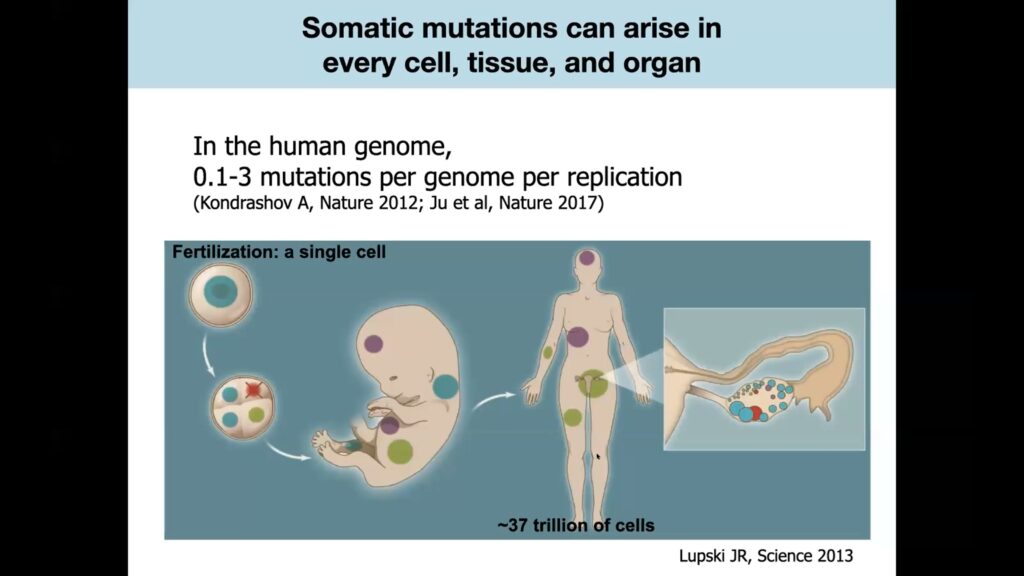


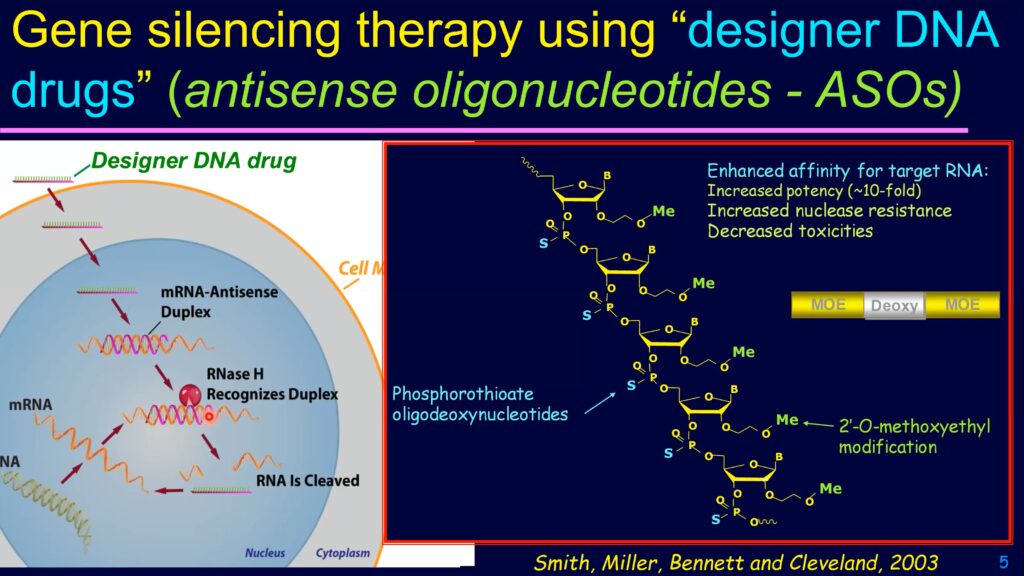
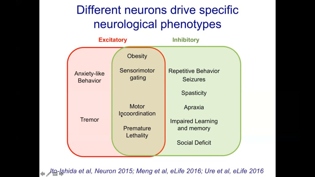
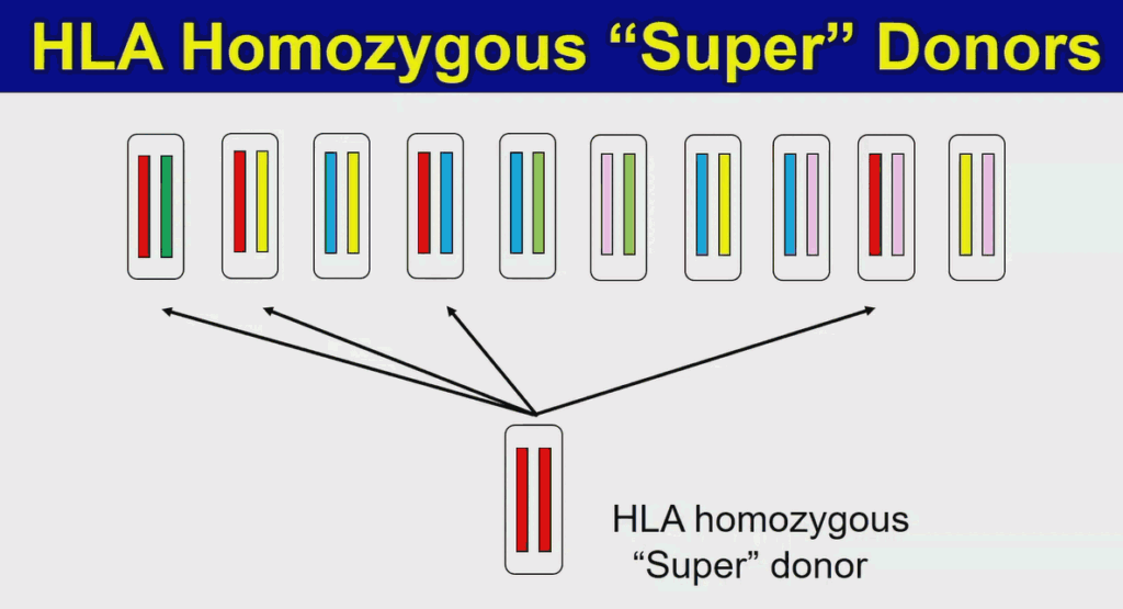
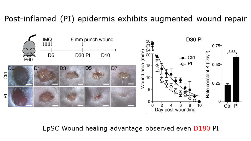
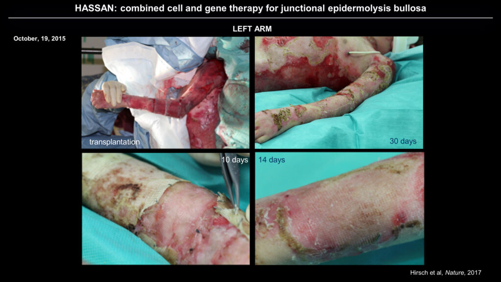
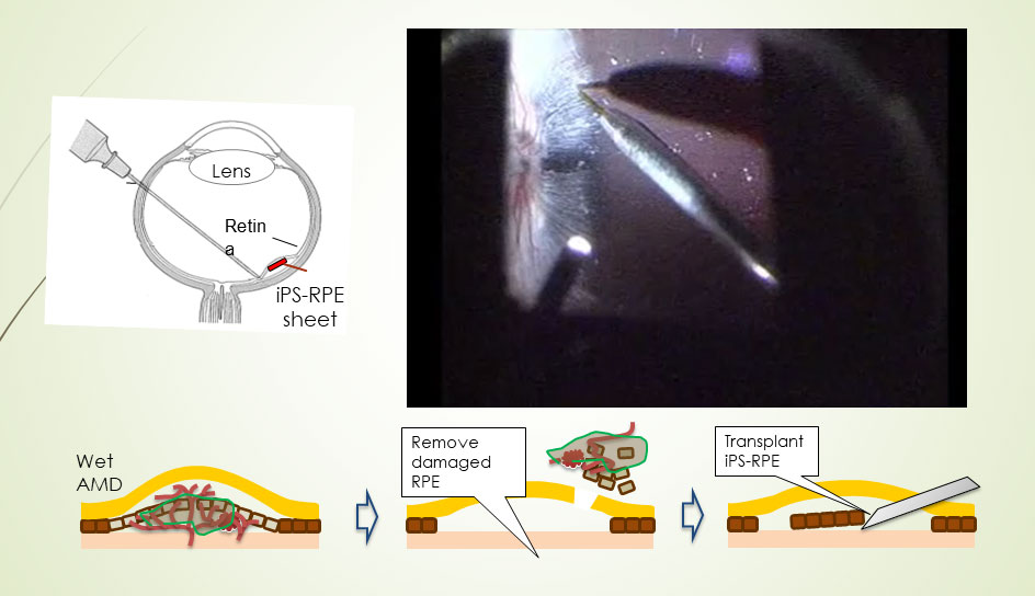
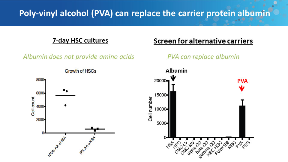
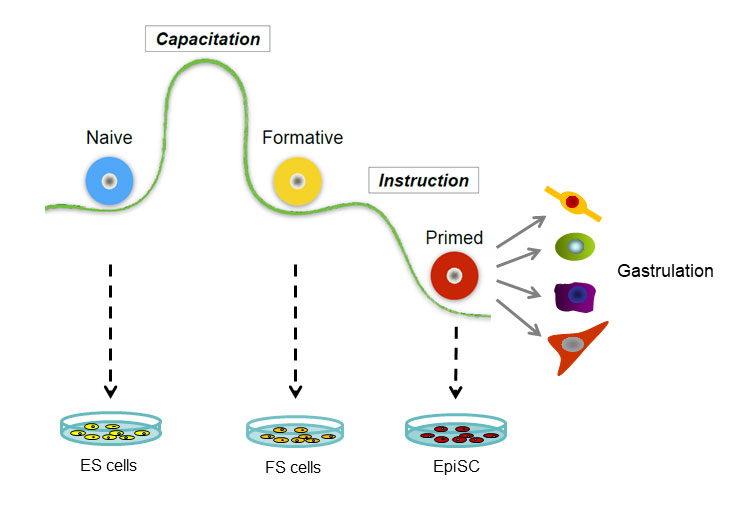


