The Innovators in Science Award Honorees are Breaking New Ground in Neuroscience: Dr. Michael Halassa’s research on AI systems could impact our perception of reality.
Published May 1, 2018
By Anni Griswold
Albert Einstein reportedly once said, “Not everything that can be counted counts, and not everything that counts can be counted.” Though the 2017 honorees of the Innovators in Science Award have plenty of countable achievements, their stories reveal a common thread — creative approaches to their work and the development of disruptive tools that transformed scientific understanding in their discipline.
Biological Underpinnings of the Mind
Michael Halassa
Michael Halassa, MD, PhD, an Early-Career Scientist Finalist, has traced the neural correlates of cognition from the thalamus to the cortex and beyond. But his interests in neurocomputational frameworks trace back even farther — to the first time he watched “The Matrix.”
As he watched the film’s characters grapple with a simulated reality, Halassa began wondering how something as intangible as the mind can perceive reality in the first place. If we were to look inside the brain, he wondered, where would we find the mind? How do we make decisions and solve problems?
“If we can understand how these functions are normally accomplished by the physical device we call the brain, then we’ll have a better understanding of how these functions go awry in conditions such as schizophrenia, autism or ADHD,” says Halassa, an Assistant Professor of Brain and Cognitive Science at Massachusetts Institute of Technology (nominated while at New York University in New York).
Computational Frameworks
Halassa abandoned the traditional tactic of studying the molecular and electrical properties of individual cells. Instead, he assembled computational frameworks that could map physical features, such as synapses, onto abstract processes such as thought. His approach revealed that the thalamus, a brain region long assumed to relay simple sensory input to the cortex, actually streams detailed instructions that allow the cortex to shift between tasks.
“From moment to moment, your brain reconfigures on the fly to perform different types of tasks. That reconfiguration is what defines things like intelligence, productivity and performance.” Glitches in this network configuration may contribute to psychiatric diseases, he says.
His findings could lead to artificial intelligence systems that display similar cognitive flexibility. Such “neuromorphic computing” could lead to a greater understanding of how we perceive reality.
Read more about Innovators in Science Award Honorees:
The Innovators in Science Award Honorees are Breaking New Ground in Neuroscience: Dr. Viviana Gradinaru’s research enables scientists to visualize neuron and cell behavior.
Published May 1, 2018
By Anni Griswold
Albert Einstein reportedly once said, “Not everything that can be counted counts, and not everything that counts can be counted.” Though the 2017 honorees of the Innovators in Science Award have plenty of countable achievements, their stories reveal a common thread — creative approaches to their work and the development of disruptive tools that transformed scientific understanding in their discipline.
Illuminating the Brain’s Circuitry
Viviana Gradinaru
As an undergraduate, Viviana Gradinaru, PhD, the Early-Career Scientist Winner, became fascinated with the underpinnings of neurodegeneration. But few tools existed to dissect the phenomenon. Undeterred, she set out to create her own.
During graduate school, Gradinaru borrowed light-sensitive proteins from algae and bacteria and introduced them to mammalian neurons. Her hope was to switch individual cells on or off in response to laser stimulation. Using this strategy, she revealed how specific brain circuits underlie locomotion, reward and sleep. One of Gradinaru’s tools, dubbed “eNpHR3.0,” is now widely used in the field of optogenetics — a field that her work helped launch.
Now an Assistant Professor of Biology and Biological Engineering at Cal Tech, Gradinaru has moved on to other tools and methods. This includes tissue-clearing techniques that render organs transparent. These see-through systems allow scientists to visualize where neurons start and stop. They also study how the cells behave along the way.
Gradinaru’s team was also among the first to introduce vectors that can shuttle genes across the blood-brain barrier with high efficiency. These genes can express colors. This allows scientists to visualize neural pathways, or they can normalize biochemical or electrical properties in a disease model.
“Developing tools and perfecting them to the level where they can work in other people’s hands,” she says, “is key to maximum impact.”
Ultimately, Gradinaru says she hopes these tools will inspire non-invasive therapies that can repair faulty brain circuits and address issues such as neurodegeneration.
Read more about Innovators in Science Award Honorees:
The Innovators in Science Award Honorees are Breaking New Ground in Neuroscience: Dr. Ben Barres inspired many with his continued efforts, in the face of his own battle with pancreatic cancer.
Published May 1, 2018
By Anni Griswold
Albert Einstein reportedly once said, “Not everything that can be counted counts, and not everything that counts can be counted.” Though the 2017 honorees of the Innovators in Science Award have plenty of countable achievements, their stories reveal a common thread — creative approaches to their work and the development of disruptive tools that transformed scientific understanding in their discipline.
Uncovering a New Role for Glia Cells: Shaping the Neural Communication Network
Ben Barres
Before Ben Barres, MD, PhD, began studying glia — cells that safeguard and anchor neurons — they were thought to play a relatively minor role in the nervous system. But Barres’ work revealed that glial cells, which far outnumber neurons, serve a more important function.
“Ben pioneered the idea that glia play a central role in sculpting the wiring diagram of our brain and are integral for maintaining circuit function throughout our lives,” said Thomas Clandinin, PhD, and professor of neurobiology at Stanford in a university press release. Clandinin was a colleague of Barres, who passed away in December 2017.
Dr. Barres inspired many with his continued efforts, in the face of his own battle with pancreatic cancer, to advance therapies for neurodegenerative disease. His obituary outlines more about his accomplished life and career.
Barres, a Senior Scientist Finalist and former Chair of Neurobiology at Stanford, began his career as a clinical neurologist. He eventually became disillusioned by the medical field’s poor understanding of neural degeneration. While reviewing pathology slides, he found that degenerating brain tissue was often surrounded by a high density of unusually shaped glial cells.
He pursued a PhD and eventually characterized three types of glial cells, revealing how they shape electrical signal transmission. He shared the tools and reagents for cloning these cells, sparking widespread interest in glial function.
Barres’ most recent work showed that rogue glial cells drive neurodegenerative disorders such as Alzheimer’s and Parkinson’s diseases, a finding he described as “the most important discovery my lab has ever made.”
Read more about Innovators in Science Award Honorees:
The Innovators in Science Award Honorees are Breaking New Ground in Neuroscience: Dr. David Julius takes a molecular approach to explore compound structures.
Published May 1, 2018
By Anni Griswold
Albert Einstein reportedly once said, “Not everything that can be counted counts, and not everything that counts can be counted.” Though the 2017 honorees of the Innovators in Science Award have plenty of countable achievements, their stories reveal a common thread — creative approaches to their work and the development of disruptive tools that transformed scientific understanding in their discipline.
Pain Relief Begins with Basic Science
David Julius
In a field as urgent and divisive as pain control, the race to market new drugs often overshadows a slower yet essential expedition: curiosity-driven science. But in David Julius’ lab at the University of California, San Francisco, curiosity has always been king.
As a graduate student in the early 1980s, Julius, a Senior Scientist Finalist, became fascinated with neurotransmitter systems. He read every paper he could find about the effects of psychoactive drugs on the nervous system. This included works by Timothy Leary and Sol Snyder. Eventually his curiosity led him to clone the serotonin receptor, a groundbreaking feat that introduced molecular biology into the field of pain research, long dominated by physiologists, pharmacologists and psychologists.
In the years since, he has taken a molecular approach to explore how plant-derived products such as capsaicin from chili peppers and menthol from mint leaves “tickle the pain pathway.” His findings have shed light on various pain receptors in the brain and uncovered ion channels that regulate sensory neurons in response to thermal or chemical stimuli.
“If any of these lead to a new pain drug, I’ll be incredibly gratified by that,” says Julius, PhD, a professor of physiology. “But in the end, these [new drugs] arise from asking basic questions about somatosensation and pain. It’s important to keep that in mind, because you never know when a basic discovery will transform an area.”
Read more about Innovators in Science Award Honorees:
On November 29, 2017, Takeda Pharmaceutical Company Limited and the New York Academy of Sciences hosted the inaugural Innovators in Science Award Symposium. The event showcased the research accomplishments of the 2017 Innovators in Science Award Honorees in neuroscience and distinguished researchers who are transforming the therapeutic landscape for neurological disease. Topics included advances in optogenetics; microglia and the role of the brain’s innate immunity in diseases including Alzheimer’s disease; advances in designing organoid systems to model complex neurodevelopmental disorders; and efforts to blend big data and systems biology to better understand the genetic drivers and heritability of autism.
Speakers
Michael Halassa, MD, PhD Massachusetts Institute of Technology
Viviana Gradinaru, PhD California Institute of Technology
David Julius, PhD University of California, San Francisco
Frederick Christian Bennett, MD Stanford University Medical School
Shigetada Nakanishi, MD, PhD Suntory Foundation for Life Sciences Bioorganic Research Institute
Rudolph E. Tanzi, PhD Harvard University
Paola Arlotta, PhD Harvard University
Daniel Geschwind, MD, PhD University of California, Los Angeles
Sponsors
Early-Career Scientist Award Honoree Lectures
Speakers
Michael Halassa, MD, PhD Massachusetts Institute of Technology
Viviana Gradinaru, PhD California Institute of Technology
Highlights
In conjunction with the prefrontal cortex, the thalamus plays an essential role in forming and applying abstract associations.
Modulating thalamic activity may have therapeutic benefit in neuropsychiatric disorders such as schizophrenia and autism.
Advances in optogenetic technologies allow researchers to map long-range pathways in the brain—an important step toward developing new therapies as well as understanding the impact of existing therapies including deep brain stimulation.
Customized viral vectors capable of delivering actuator genes to the brain via the circulation are a potential means for non-invasive neural modulation.
Rethinking the Thalamus
Michael Halassa opened the Symposium with the first of two presentations from early-career scientists whose findings hold promise as the basis of future therapies.
Halassa’s work challenges conventional beliefs about the role of the thalamus, suggesting that rather than a simple relay for delivering sensory information to the brain’s cortex, the region is a “superhighway” that facilitates the application of abstract associations formed in the prefrontal cortex. The ability to capture information about the outside world, organize and preserve it hierarchically, and apply those associations in real-time form the basis not only of perception, but of higher-order skills such as prediction and storytelling. Halassa explained that dysfunction in these uniquely human mechanisms underlie schizophrenia, autism, and other neuropsychiatric disorders. Understanding the role of the thalamus in these processes may lead to new therapies and novel approaches to cognitive enhancement.
While the thalamus is, indeed, a sensory relay, only a small portion of the structure is responsible for these functions, while most play no role in sensory processing, Halassa explained. The mediodorsal thalamus (MD) is involved in working memory and other cognitive tasks, and is strongly connected to the prefrontal cortex, the seat of executive functioning and decision-making. Experiments in mice reveal that the thalamus is a critical conduit for interaction between sensory information entering the thalamus and abstract thoughts and associations in the prefrontal cortex.
Mice were trained to associate sensory stimuli—in this case, two distinct sounds—with a specific task directive. One sound indicated that the mouse should respond to a visual cue, the other indicated attention to an auditory cue. Using optogenetics to visualize and modulate regions of the thalamus and prefrontal cortex, the researchers discovered that the process of turning a sensory stimulus into a behavioral rule relies on input from the prefrontal cortex. When either the prefrontal cortex or the thalamus is inhibited, mice can no longer direct their attention accordingly— the ability to tap into the association and follow the rule is impaired.
Mice do not respond to sound cues associated with task rules outside of the experimental environment.
Patterns of neuronal activity show that while the prefrontal cortex is critical for forming associations, the thalamus sustains them. “Signals in the thalamus unlock associations and encode context, which allows the brain to be flexible depending on the circumstances,” said Halassa. When mice were given the same sound cues outside the experimental environment, they make no association. “Different stimuli have different meanings depending on the context, and we see this in everyday life—if you see a red light while you’re driving, you stop. If you see a red light at a dance club, you don’t,” he explained.
Pharmacologic enhancement or dampening of thalamic function may hold promise as a means to address the dysfunctions in contextualization and abstract associations seen in schizophrenia and other psychiatric disorders.
New Approaches to Visualizing and Controlling Brain Circuity
Optogenetic tools have allowed researchers to visualize and control the complex circuitry of the brain. Despite these advances, much remains unknown about the neural paths that influence behavior and brain function in health and disease. Viviana Gradinaru, winner of the Early-Career Scientist Innovators in Science Award, discussed cutting-edge refinements to optogenetic techniques that, combined with modern tissue-clearing methods, enable the type of detailed neural mapping critical to harnessing the brain’s circuitry to treat disease.
Microbial opsins, the light-sensitive proteins that make optogenetics possible, can often be adapted to function in mammalian cells with relative ease. “It’s remarkable, when you think about what a big ask it is for these sequences to function the way we want them to,” said Gradinaru, noting that in their native hosts, such proteins enable entirely different functions, such as locomotion. As optogenetics applications have expanded, however, Gradinaru and others have tweaked the sequences and packaging of these workhorse proteins to increase precision, tolerability, functionality, and to devise new means of delivery.
Gradinaru is developing tools capable of illuminating the brain’s long-range pathways. Mapping the fine projections of the brain poses significant challenges: slicing the tissue can compromise reconstruction of the pathways, and the poor optical properties of lipid-rich brain tissue make imaging difficult. In a significant step toward solving these problems, Gradinaru revived the century-old technique of tissue clearing, adding modern improvements that dissolve light-scattering lipids while locking proteins and nucleic acids in place within a hydrogel matrix that preserves structure. The resulting tissue is both transparent and capable of retaining colored labels. Used in tandem with a new class of optogenetic tools, this tissue-clearing technique enables researchers to track individual axons, even through tightly packed bundles.
New visualization protocols combining whole body tissue clearing with custom viral vectors that deliver genes systemically and allow for cell type-specific expression facilitate high-resolution, intact neural circuit mapping.
Viral vectors are a common means of introducing labels, sensors, and actuators into the brain, typically via intracranial injection, a method that is invasive and often results in non-homogeneous expression over limited tissue volume. “But what if we could use the vasculature to deliver labels brain-wide, and devise a way to control the density of the labels?” Gradinaru asked, noting that the major hurdle to delivering opsins to the brain is finding a vector capable of circumventing the blood-brain barrier. Using directed evolution, Gradinaru and her team at Caltech engineered a strain of a common viral vector. The adeno-associated virus is capable of passing through the blood-brain barrier and delivering a customized package of protein sequences and gene-regulatory elements that allow researchers to refine a target and restrict expression to certain cell types. It is also possible to limit expression within a cell type, which reduces the density of the label and allows for more precise observations. “We can systemically deliver various colors and direct their expression in the brain, then clear the tissue and reconstruct the morphology to see the long-range projection pathways,” said Gradinaru. Looking to the future, she envisions systemically delivered genes (e.g. actuators or editing tools) as a non-invasive method of modulating neuron biochemistry and activity.
David Julius, PhD University of California, San Francisco
Frederick Christian Bennett, MD Stanford University Medical School
Shigetada Nakanishi, MD, PhD Suntory Foundation for Life Sciences Bioorganic Research Institute
Highlights
Studies of mechanisms by which natural compounds such as capsaicin and menthol modulate temperature-sensitive ion channels in the peripheral nervous system are leading to greater understanding of pain perception.
Recent experiments reveal that while microglia lose expression of highly specific, signature genes in culture, reintroduction to the CNS environment induces their re-expression and a return to functionality, hinting at the possibility for microglial transplant as a therapy for diseases including Alzheimer’s.
Discoveries over the past three decades have revolutionized our understanding of neurotransmitter-receptor interactions and the role of these pathways in health and disease.
Probing the Pain Pathways
David Julius began a session of presentations by senior scientist honorees with a discussion of pain pathways—crucial protective mechanisms that often go awry, resulting in prolonged suffering and disability. According to Julius, pain is the primary reason patients seek medical help, and in the United States alone, up to 100 million people suffer some form of persistent pain.
Elucidating the mechanisms of pain—particularly those that drive the shift from acute pain, which is purposeful, to chronic pain, which can persist long after injury subsides—is key to understanding how to address and prevent persistent pain syndromes. A family of cation ion channels, the Transition Receptor Potential or TRP channels, play major roles in sensory physiology, facilitating detection of a wide range of both endogenous and exogenous stimuli including temperature, pressure, chemical irritation, and inflammation. Julius discussed his lab’s discoveries about the structure, functionality, and modulation of a subset of TRP channels tied to temperature and/or chemical sensitivity: TRPV1, TRPM8, and TRPA1.
Julius and his collaborators turned to nature for both inspiration and information to guide their research, focusing on several of the plant kingdom’s natural defense mechanisms: the potent chemicals capsaicin, which produces the sensation of heat in chili peppers; menthol, the cooling agent in mint oils; and thiosulfinates, pungent compounds found in onion and garlic. These “homegrown chemical warfare agents” ward off predators by producing an acute pain response, and it is the impact of these chemicals on TRP channels that form the basis of research that is answering foundational questions about the mechanisms and structure of these complex cellular sensors and their role in pain signaling.
Heat, Cold, Pain, Pressure
Two of the ion channels studied, TRPV1 and TRPM8, are temperature sensors, with the former activated by heat and the latter by cold. Experiments show that TRPV1 can be activated chemically by capsaicin, but also by changes in ambient temperature. “The channel is quiet at body temperature, but at about 43 degrees, it strongly activates,” said Julius. “Interestingly, this is the psychophysical threshold at which most of us discriminate between an innocuously warm object versus one that’s noxiously hot.”
TRPV1 and TRPM8 are triggered by changes in ambient temperature.
Conversely, TRPM8 is activated at temperatures below 25 degrees, as well as by exposure to menthol. In a physiologic twist, actual sensations of cold and heat and chemical compounds that mimic cold and heat are perceived similarly by the peripheral sensory nervous system. Further studies reinforce that these proteins are intrinsically temperature-sensitive, and serve as molecular thermometers essential for detecting and responding to temperature shifts. To wit: mice bred without a TRPM8 gene are unable to distinguish between a warm compartment and an uncomfortably cold one.
TRPV1 can also be modulated by components of the body’s “inflammatory soup,” explained Julius, noting that while some modulators, including extracellular protons and some lipids, bind directly to the channel and lower its threshold for detecting heat, other modulators, such as prostaglandins and peptides, act on their own receptors yet have the same threshold-lowering effect on TRPV1. “Understanding how these allosteric components directly or indirectly act on the receptor to change its threshold is a step toward developing drugs that interfere with the ability of these modulators to over-sensitize the channel and contribute to persistent pain syndromes,” Julius said.
Modeling the Channel
A lack of high-resolution atomic structural information has hindered researchers’ efforts to fully understand the functionality and modulation of TRP channels. Julius described how cryo-electron microscopy has yielded the first three-dimensional models of TRP channels and allowed researchers to observe the channels’ gating mechanisms in response to multiple stimuli. Imaging reveals that TRP channels have two operable gates which control ion flow, with some modulators acting specifically on one gate or the other. Julius believes this structure contributes to the channels’ sensitivity and efficiency as sensors. “This suggests that part of the reason TRP channels function so beautifully as polymodal signal detectors is that this two-gate mechanism allows for very rich physiologic modulation,” he said.
More recently, Julius and his collaborators have begun to study TRP channels embedded in lipid nanodiscs, which mimic the native environment of the body. Doing so allows them to visualize the precise location where molecules bind to the channel, including toxins and small molecules—an important step in designing next-generation analgesic drugs to modulate the activity of these channels.
Origin, Environment, and Microglial Identity
Frederick Christian Bennett addressed the Symposium on behalf of the late Ben Barres, Senior Scientist Award finalist. Bennett began with an homage to his mentor, highlighting the pioneering neurologist and neuroscientist’s contributions to discovering the dual role of astrocytes—as critical facilitators of synapse formation and functioning in health, and as highly reactive synapse-and neuronal destroyers in certain stress or disease states. “Glial cells and brain function are intimately intertwined in complicated ways we are just beginning to understand, and which hold enormous potential as treatment targets,” Bennett said, transitioning into a discussion of his own research, which also centers on a type of glial cell—microglia.
Microglia are macrophages often referred to as the resident immune cells of the central nervous system. Arising from the yolk sac early in fetal development and quickly sequestered inside the brain, microglia have an origin unlike other hematopoietic cells in the body, which arise in the bone marrow and differentiate throughout the body. Like astrocytes, microglia are dynamic cells capable of changing state based on context and environment, and much the way astrocytes assume a reactive state in diseases such as Alzheimer’s, ALS, and multiple sclerosis, microglia too become dyshomeostatic in disease.
Until recently, in-depth studies of microglial cell function have been complicated by an inability to distinguish them from other macrophages in the brain. However, microglia express a series of signature genes that differ from those expressed by other macrophages. Antibody-based techniques that bind to TMEM119, a protein highly specific to microglia, have yielded new insights into these unique cells. By tracking expression of TMEM119, Bennett is investigating how the CNS environment impacts microglial function and gene expression, and whether microglial cell transplant may be a viable treatment strategy for brain diseases.
Bone marrow transplants are already used to treat certain disorders that impact brain function, with the goal of introducing hematopoietic cells that will infiltrate the brain and differentiate into healthy microglial cells to replace those malfunctioning due to disease or mutation. Yet, studies show that while stem cells do enter the brain and differentiate into cells that are “microglia-like,” they are not true microglia and do not express TMEM119 or other signature microglial genes. The concept of a true microglial cell transplant is one Bennett deems “provocative,” and despite the early stage of the research in animal models, promising.
Bennett’s research shows that the unique ontogeny of microglia, along with elements of the CNS environment, have a profound influence on the cells’ identity and function. Experiments with cultured mouse microglia reveal that these cells lose all expression of signature genes, including TMEM119, when removed from their native environment. Conversely, in a surprising display of plasticity, cultured microglia resume normal gene expression when returned to the CNS. “Signals in the brain are sufficient to sustain, induce, and re-induce microglial identity,” Bennett said. “We didn’t know that it was possible to take cells out of the brain, reintroduce them, and have them recover their homeostatic state.” He explained that such findings may upend notions about the state of microglia in diseases such as Alzheimer’s. For example, it has been posited that in a CNS environment rendered abnormal by disease, microglia may enter an irreversibly neurodegenerative state. The ability of microglial cells to change gene expression and functionality based on their environment suggests that perhaps changes to the CNS in brain disease may restore microglial function.
Some of the most fundamental insights about the molecular mechanisms that facilitate communication between neurons have emerged from the work of Shigetada Nakanishi. In a career spanning five decades and counting, Nakanishi has discovered and elucidated the roles for multiple families of neurotransmitters, including the ubiquitous excitatory amino acid glutamate, which is critical for neural plasticity, learning, and memory tasks. In excess, glutamate is also implicated in neurodegenerative disorders.
Glutamate receptors are categorized as either ionotropic, meaning they directly and rapidly control ion flow by opening or closing pores in the neuronal cell membrane; or metabotropic, meaning they act on signaling pathways within the nerve cell. These characterizations were discovered using a technique Nakanishi pioneered to circumvent a major hurdle facing early studies of neurotransmitters. Namely, the substances were simple to extract and study, but the receptors, buried deep within cell membranes, eluded extraction and cloning. Nakanishi’s method, which used electrophysiology to measure expression of glutamate receptors in Xenopus oocyte cell membranes, yielded the molecular identities of more than a dozen subtypes of ionotropic glutamate receptors and eight metabotropic subtypes. In the decades that followed, he has demonstrated the role of various receptors in facilitating key brain functions and behaviors, including responding to reward-seeking or aversive stimuli and discriminating between the presence or absence of light.
Nakanishi described experiments targeting the dopamine pathways that control reward-based learning in the basal ganglia to better understand how these two morphologically similar but functionally distinct pathways modulate reward-seeking and aversive reactions. Rewarding or aversive stimuli enter the nucleus accumbens via glutamate transmission, and these inputs are further transmitted along two parallel pathways—the direct pathway, characterized by neurons that express excitatory D1 dopamine receptors, and the indirect pathway, packed with neurons that express inhibitory D2 receptors. “These pathways modulate behavior through dopamine transmission, but in opposite ways,” said Nakanishi.
Glutamate receptors facilitate transmission along separate and opposing pathways in both the retinal and the striatal neural networks. The result is the ability to discriminate between light and darkness, and to interpret stimuli as either rewarding or aversive.
Nakanishi and his collaborators used tetanus toxin to selectively and reversibly block each transmission pathway, discovering that the direct pathway (D1) increases dopamine levels and is responsible for processing rewarding stimuli, while the indirect (D2) pathway processes aversive stimuli and reduces dopamine transmission. More advanced studies of this phenomenon revealed that rewarding and aversive stimuli are converted to activation of excitatory D1 receptors and suppression of inhibitory D2 receptors, respectively; this conversion commonly activates the cAMP-protein kinase A signaling cascade and in turn, stimulates glutamate transmission in a stimulus-dependent, pathway-specific manner.
A similarly structured neural transmission network governs the discrimination between light and dark in the retinal network, explained Nakanishi. Here too, stimulation of highly specific and opposing receptors—AMPA and mGLUR6, both glutamate receptor subtypes—results in the ability to distinguish light and dark signals.
He noted that imbalances in neurotransmitters and functioning of their receptors are implicated in both organic brain diseases and psychiatric diseases, including depression. Illuminating complex neurotransmitter-receptor interactions is critical for understanding healthy brain function as well as the origin and progression of disease, and is fundamental to drug development, as many drugs act directly on receptors.
Amyloid deposition begins 10–15 years before patients present with symptoms of Alzheimer’s disease, indicating a need for early identification and treatment of at-risk patients.
Three-dimensional neural cell cultures that recapitulate Alzheimer’s disease in a dish are facilitating the identification of compounds that prevent or slow the formation of plaques and tangles.
Beta amyloid is a powerful anti-microbial peptide, and emerging research points to the protein’s immune function as a possible factor in “seeding” plaques that can ultimately lead to Alzheimer’s.
From Rudolph Tanzi’s perspective, the five million patients in the United States with Alzheimer’s are only the tip of the iceberg when it comes to the true scope of the disease. “Plaques begin to form 10–15 years before patients present with symptoms, so that’s potentially 20 or 25 million people who are on the path toward a diagnosis,” said Tanzi, emphasizing that unlike heart disease or cancer, Alzheimer’s disease (AD) requires patients to present with symptoms before treatment begins. “Imagine if we waited until cancer was symptomatic before we treated it?” he said. “Trying to treat plaques once someone has dementia is like trying to put out a forest fire by blowing out the match.”
Tanzi is a co-discoverer of the early-onset familial Alzheimer’s genes, and 30 years after these initial discoveries, the field of genomics has been transformed through advanced sequencing techniques. These techniques allow researchers to identify areas of the genome where AD genes reside as well as mutations that, when studied in mice and in cultured human nerve cells, are leading to next-generation therapeutics aimed at preventing or halting disease progression. Over the past decade, Tanzi and others have built an ever-growing list of more than 200 AD-related mutations impacting one or more aspects of the “trilogy of pathology” underlying Alzheimer’s—amyloid plaques, tangles, and neuroinflammation.
Attempts to recreate Alzheimer’s in mouse models has done little to shed light on the interplay of these factors in causing AD, but in recent years, the advent of brain organoids—human stem cell-derived neural culture systems—has facilitated the creation of what Tanzi terms “Alzheimer’s in a dish.” For the first time, researchers can observe a disease progression that typically takes more than a decade in a matter of weeks. Tanzi reported that by 7 weeks, cells expressing APP or presenilin mutations develop both plaques and tangles, presenting an unprecedented opportunity to test the results of various interventions on the disease process.
Human neural cultures of Alzheimer’s disease, or “Alzheimer’s in a dish” are an efficient, effective method for screening compounds that may impact AD pathogenesis, such as those that inhibit the formation of neurofibrillary tangles.
Blocking amyloid deposition through beta or gamma secretase inhibitors successfully reduces the incidence of plaques but also, notably, blocks the formation of tangles—firm proof of the long-debated “amyloid hypothesis,” or the notion that amyloid itself is the driver of AD. Tanzi and his collaborators have also used this model to test compounds that effectively block tangles, identifying 30 FDA-approved drugs that inhibit tangle formation, and another 8 that reduce amyloid deposition, and thus block tangles.
Neuroinflammation is the least-studied factor in AD pathogenesis, yet it warrants greater attention. Tanzi explained that brains that are resilient to Alzheimer’s—those replete with plaques and tangles that never cause dementia—are united by a common feature: a lack of inflammation. “You can have lots of plaque and tangles, but if the brain’s immune system doesn’t react to them with gliosis, you can escape dementia,” he said. Genes including TREM2 and CD33 are implicated in the neuroinflammatory process by regulating the activity of microglial cells. Mutations or knockouts of these genes can impair microglial plaque clearance as well as promote pro-inflammatory changes in these critical immune cells.
Tanzi concluded by describing recent studies that reveal a surprising dual role for beta amyloid, not only as a pathogenic protein in AD but also as an anti-microbial peptide and an essential component of the brain’s innate immunity. The tendency of amyloid beta to form plaques, viewed as inherently pathological in AD, is a powerful protective mechanism when viewed in an immune context. The protein binds to carbohydrates on microbial surfaces, agglutinates, and ultimately traps invading pathogens within a plaque. When HSV1 and Salmonella are introduced into mouse brains, beta amyloid deposition skyrockets, forming plaques in as little as 24 hours. “It’s still early days for this research, but it may be that microbes are seeding amyloid deposition in the brain. This is an entirely different way to look at the origin of Alzheimer’s,” Tanzi said.
AJNR Am J Neuroradiol. 2008 Jan;29(1):18-22. Epub 2007 Oct 9.
Neuropsychiatric Diseases
Speakers
Paola Arlotta Harvard University
Daniel H. Geschwind University of California, Los Angeles
Highlights
Long-cultured brain organoids differentiate into many cells types, including light-sensitive retinal cells and neurons with dendritic spines.
Organoids can be used to model highly penetrant single mutations that impact neurodevelopment, and will soon be a viable method for creating models of polygenic disorders in culture.
Autism is one of the most heterogeneous neurodevelopmental disorders, encompassing a wide range of phenotypes and a large and rapidly growing number of associated genetic mutations.
Analyses incorporating genetic, transcriptomic, and phenotypic data are revealing patterns of gene expression in autism, as well as linkages to neuropsychiatric disorders including schizophrenia and bipolar disorder.
Building the Brain: From Embryo to Organoid
“How can we model hugely complex diseases like autism, schizophrenia, and other disorders that impact aspects of personality and behavior that are uniquely human?” asked Paola Arlotta, describing the difficulty of unraveling the mysteries of neurodevelopmental disorders. “If we want to look at the impact of the whole genome on disease, we can’t use an animal model—we need a human model,” she said. Arlotta’s lab builds brain organoids—organ models cultured from embryonic or pluripotent stem cells—to understand both normal and atypical brain development in unprecedented detail.
Organoids are a relatively new but promising model for investigating brain development and neurodevelopmental disorders, but until recently, the lifespan of an organoid was measured in weeks. Thanks to modifications in the protocols, Arlotta’s lab is building brain organoids that grow for nine months or longer, maturing and diversifying into an array of cell types and providing new insights into the human neurodevelopmental process.
Much of this insight has been gleaned through single-cell studies of more than 80,000 cells taken from 30 organoids and examined for cell type classification, patterns of gene expression, and physical characteristics. Arlotta and her primary collaborator, postdoctoral fellow Giorgia Quadrato, reported the discovery of a surprising array of cell types, including astrocytes, radial glia, excitatory and inhibitory neurons, retinal cells—including photoreceptor-like cells that respond to light—and progenitors of the cerebral cortex. While little is known about the patterns of gene expression in cells of the fetal human brain, comparisons of the various cell types identified within the organoids with the limited existing data show high correlation between the cells in the organoid and endogenous fetal brain cells. “With no instruction from the outside, these organoids self-organize and mimic the process of human brain development, including differentiating into these various cell lineages,” Arlotta said. “It really speaks to the power of what’s encoded in the genome.”
Cell samples from 8-month-old organoids strongly recapitulate both cell type diversity and patterns of gene expression seen in endogenous fetal brain cells.
In a subsequent set of findings that stunned both Arlotta and her collaborators, cells from an 8-month-old organoid were shown through electron microscopy to contain dendritic spines, indicating a level of cell maturity difficult to achieve in culture. Psychiatric diseases including schizophrenia are tied to synaptic dysfunction and errors in dendritic pruning, thus the ability to grow cells that form true spines in a dish may present new avenues for exploring genetic drivers of these diseases as well as potential treatments.
“We are heading toward a future where we can engineer highly penetrant mutations into pluripotent stem cells, grow an organoid, and use single-cell sequencing to understand what cell types and pathways are affected by that mutation,” Arlotta said. Noting that most neurodevelopmental diseases are polygenic, she also emphasized the potential for creating chimeric organoids that express different mutations and can be grown from human cells with any genetic background. “An individual’s genomic background is fundamental in controlling the outcome of a mutation, and we’ll soon be able to use organoids to observe how the genome modulates the effect of a certain mutation,” she said.
Autism: Genes, Heritability, and Risk
Daniel Geschwind delivered the final research presentation of the Symposium—a brief but intense dive into efforts to discover the genetics of perhaps the most heterogeneous neurobiological disorder: autism.
Geschwind applies a systems biology approach to the challenge of identifying causal genetics and heritable risk of a disorder that, unlike neurological diseases, evidences no physical manifestations of pathology. Autism is diagnosed based on observations of behavior and evidence of deficits in social interaction and communication, and the disorder encompasses a wide range of phenotypes. Geschwind is building data sets including genetic, transcriptomic, and phenotypic information in an attempt to uncover and understand the linkages between the many manifestations of autism, as well as its connection to other neurodevelopmental and neuropsychiatric disorders.
Geschwind explained that autism is largely driven by common genetic variants, along with some rare de novo mutations, yet no specific gene accounts for more than 1%–2% of cases. A decade ago, fewer than 10 genes were associated with autism, but due to a surge of research interest in the disorder, which affects 1 in 68 children in the United States, more than 200 autism-associated genes have now been identified. Geschwind expects that number to quickly rise to at least 1,000. Due in part to this extreme genetic variability, he and his collaborators are probing the transcriptome in search of patterns of gene expression and regulation that may transcend the genetic and phenotypic heterogeneity of autism.
Comparisons of cortical tissue in autistic and neurotypical brains reveal distinct differences in patterns of co-expression in genes that drive corticogenesis and synaptic function “Most of these genes are expressed at high levels in fetal development, which tells us that a good deal of autism risk that we can identify is occurring during fetal brain development, and that’s quite important,” said Geschwind. Notably, brains of people diagnosed with autism are characterized by downregulation of genes associated with synaptogenesis, and upregulation of genes that regulate microglia and astrocyte activity. As glia are critical for synaptic plasticity, Geschwind believes that this abnormal upregulation of their activity may promote a dyshomeostatic environment that impairs synaptic function in the autistic brain. “It’s also possible that what we see as an upregulation may just be the failure of the neurotypical downregulation of these genes in the first decade of life, so it’s possible that there’s a treatment window there,” Geschwind said.
Advances in sequencing and a surge of interest in ASD have led to the identification of more than 200 gene mutations associated with autism, none of which accounts for more than one percent of cases and many of which have strong pleiotropy.
Transcriptomic comparisons between autism and psychiatric disorders including schizophrenia, bipolar disorder, and depression provide additional insights regarding the correlation of disorders that are clinically distinct yet show strong co-heritability. Geschwind described overlaps in gene expression in autism and schizophrenia, as well as in schizophrenia and bipolar disorder—similarities that lay, in part, in the common upregulation of astrocytes and/or microglia in these disorders.
Ascertaining the impact of specific autism-association mutations on neurodevelopment requires a new generation of tools that faithfully recreate in vivo neurodevelopment in vitro or in silico. Geschwind described how machine learning algorithms and 3D neural cell cultures—brain organoids—are advancing the quest to understand the interplay of these complex factors.
Dr. Richard Gilbertson discusses his inspiration and the latest advances in pediatric cancer research.
Published January 8, 2018
By Marie Gentile and Richard Birchard
Dr. Richard Gilbertson
Richard Gilbertson, MD, PhD, Li Ka Shing Chair of Oncology and director of the Cancer Research UK Cambridge Centre, did not initially set out for a career in pediatric cancer — the leading cause of death by disease past infancy for children and adolescents in the United States and Europe.
He “somewhat randomly,” as he says, chose to do his second-year research project on medulloblastoma, the most common malignant brain tumor in children. He was inspired early on by a caring mentor who went above and beyond in attention and enthusiasm and was further determined to pursue this path while getting to know the family of a child with brain cancer.
“One day I went onto the ward, and it was very dark, and all the curtains were closed, and I was told that this child was dying. After inquiring about available treatments, I was told there was nothing to be done. I was incredibly angry with the system that wasn’t able to offer a child a curative treatment.”
Deeply affected by this child’s death, when a friend and fellow medical student challenged him to produce a 15% reduction in mortality of any disease over beers at a pub, Dr. Gilbertson made it his career goal to “produce a 15% reduction in mortality, at least of medulloblastoma in pediatric cancer.”
Discoveries in Medulloblastoma
To that end, Dr. Gilbertson and his lab have made some profound discoveries in medulloblastoma. During the 1980s, medulloblastoma was considered a single disease, with a singular treatment, but “we’ve demonstrated that it is multiple diseases, and those diseases actually have different origins in the nervous system from very specific cell types, and they behave differently.”
This understanding has allowed treatments to be tailored to disease type, resulting in a reduction in the use of radiation therapy, the introduction of new treatments that target the signaling pathways of some forms of medulloblastoma, and insights into other brain tumors including Ependymoma and choroid plexus carcinoma.
His latest research is driven by the question of why cancer is so much less prevalent in children than expected, given that as they grow they have a large burden of cellular proliferation.
“Whereas one in two adults will get cancer eventually, only one in 600 children will, and the math doesn’t add up because children are growing faster than at any other point in their lives,” says Gilbertson.
Understanding the Mechanisms of Cancer Protection
Researchers have long suspected that children’s tissue provides protection against cancer to accommodate this growth, but they lacked definitive evidence or a mechanism for how this works. In a landmark paper published in Cell, Dr. Gilbertson’s lab mapped the functions of cells in numerous organs across the lifetime of mice and introduced tumor-inducing mutations to those cells.
They found that neonatal mouse cells are less likely to undergo tumorigenic transformation compared to adult cells with the same stem cell capacity, supporting the hypothesis that neonatal cells are somehow resistant to forming tumors — extrapolating to humans, this may explain why cancer rates are lower in children than adults.
Understanding the mechanism of this cancer protection has the potential to lead to better treatments not only for pediatric cancers, but adult cancers as well. “That’s critically important because if I can understand (how pediatric cells are protected from cancer), and then we can reactivate that in adult tissues, you’d have a very potent cancer preventative. If we could reactivate the mechanism in pediatric cells to allow them to grow and repair, but not cause cancer — imagine what we could do in adults. You could actually reactivate that pharmacologically with a medicine.”
Dr. Gilbertson is adamant about the need to develop innovative treatments that are proactive and integrated.
“My passion is to see cancers diagnosed as early as possible. Obviously, if you diagnose a cancer earlier, and this is particularly important for children, the required treatment is much less intense. The heroes of future cancer care may not so much be the life scientists, but the physicists, chemists, engineers, and mathematicians. They will be the people who generate innovative and inexpensive devices to detect cancer in its very earliest stages across the population,” he says.
The Need for International Collaboration
Dr. Gilbertson presented his groundbreaking work during the opening Keynote Lecture at the 2018 Sohn Conference: Accelerating Translation of Pediatric Cancer Research, which brought together the leaders in the field of pediatric oncology, and allowed interactions between more established scientists and clinicians with the next generation of graduate students, post-docs, and other young investigators from around the world. This was particularly exciting because due to the rarity of pediatric cancer, clinical trials to develop new treatments require international collaboration. “This disease is life threatening, there’s an imperative to do the best possible research.”
Many promising strategies for promoting neuroregeneration have emerged in the past few years, but a further research push is needed for these ideas to be translated into therapies for neurodegenerative diseases.
On June 13–14, a symposium presented by Eli Lilly and Company and The New York Academy of Sciences brought together academic and industry researchers working on multiple neurodegenerative diseases as well as clinicians and government stakeholders to discuss cutting edge basic and clinical research on neuroregeneration and neurorestoration. Topics included neuronal plasticity, inflammation, glial cell function, autophagy, and mitochondrial function, as well as analysis of recent drug development failures and how to move forward from them.
Speakers
Benedikt Berninger, PhD, University Medical Center Johannes Gutenberg University Mainz, Germany
Graham Collingridge, PhD, University of Toronto
Ana Maria Cuervo, MD, PhD, Albert Einstein College of Medicine
Valina Dawson, PhD, Johns Hopkins School of Medicine
Roman Giger, PhD, University of Michigan
Steven Goldman, MD, PhD, University of Rochester Medical Center
Eric Karran, PhD, AbbVie
Arthur Konnerth, PhD, Technical University of Munich, Germany
Guo-li Ming, MD, PhD, Johns Hopskins School of Medicine
David Rowitch, MD, PhD, ScD, University of Cambridge and University of California, San Fransisco
Amar Sahay, PhD, Massachusetts General Hospital
Reisa A. Sperling, MD, MMSc, Brighman and Women’s Hospital
James Surmeier, PhD, Northwestern University
Richard Tsien, DPhil, New York University, Longone Medical Center
Jeffrey Macklis, Harvard University
Mark Mattson, National Institute of Aging
Clive Svendsen, Cedars-Sinai Medical Center
Michael Sofroniew, David Geffen School of Medicine, UCLA
Michael J. O’Neill, Eli Lilly and Company
Presented By
Meeting Reports
Meeting Reports
Astrocytes in CNS Repair; Disease-Modifying Therapies in the Pipeline
Speakers
Eric Karran AbbVie
Michael V. Sofroniew David Geffen School of Medicine, University of California, Los Angeles
Highlights
Astrocyte scar formation is not detrimental to neuronal regeneration and repair after injury but is in fact critical to the healing process.
The clinical pipeline in Alzheimer’s disease is dominated by amyloid beta-targeting compounds, despite the fact that the approach has not been successful to date.
Astrocytes in CNS Repair
In his keynote talk, Michael V. Sofroniew of the University of California, Los Angeles, described 25 years of work on the overlooked and misunderstood role of astrocytes in the central nervous system (CNS).
These glial cells were discovered in the 19th century, and researchers widely believed that their activation after injury—which often results in scar formation around the lesion—detrimentally affects recovery. “But one has to ask, why would nature conserve this response to injury across all mammalian species if it were purely detrimental?” Sofroniew said.
Astrocytes can play fundamentally different roles in the CNS. In healthy tissue, they help synapses take up and release neurotransmitters and other factors, and help maintain neuronal energy balance and blood flow in surrounding tissue. Their activation in response to damage differs depending on whether recovery requires neurons to grow through lesioned tissue or through intact neural tissue.
Two different phenotypes of reactive astrocytes occur after injury.
Astrocytes responding to injury exist in different phenotypes: a hypertrophic reactive form interacts with neural cells, and a scar-forming reactive form interacts with non-neuronal inflammatory and fibrotic cells. Researchers are just beginning to define the function of hypertrophic astrocytes, but Sofroniew and his colleagues hypothesize that they represent a beneficial gain of function—helping injured neurons make new synapses and reorganize damaged circuits. Much remains to be learned about this process, he said.
Ongoing research from Sofroniew’s lab suggests that scar-forming astrocytes have a different, also beneficial function: recruiting inflammatory cells into the tissue, regulating their activity, and restricting their spread outside the lesion. Inflammation is crucial for getting rid of damaged cells, but too much of it damages surrounding intact tissue.
When neural tissue is injured, astrocytes recruit cells to scavenge damaged tissue. Somehow, astrocytes sense where the border between damaged and healthy tissue should be and wall off the injury with scar tissue. Sofroniew and others have shown that disrupting scar formation causes neurons in surrounding tissue to die.
Entrenched dogma in the field, however, says that astrocyte scar formation prevents axon regeneration. Twenty years ago, Sofroniew’s lab first tested whether disrupting scar formation in mice would spur injured axons to spontaneously regenerate. Their results showed that it didn’t, but the findings went against current dogma so the team never published them. When a researcher interested in the question joined the lab recently, they began exploring the question again, using two mouse models with mutations that prevent scar format.
After a spinal cord injury, sensory axons stimulated with growth factors can regrow despite astrocyte scar formation.
They showed that axons in three different types of CNS tracts failed to regrow in the mutant mice. Both astrocytes in lesions, along with other, non-astrocyte cells, all produced a variety of molecules both promoting and inhibiting axonal growth, underscoring the multi-component nature of regeneration. And axons that received appropriate stimulatory molecules “grow happily across astrocyte scars,” he said. The group is now confirming the result in additional types of CNS tracts. Sofroniew concluded that astrocyte reactivity and scar formation are not forms of astrocyte dysfunction, but adaptive functions critical for CNS repair and regeneration after injury.
Disease-Modifying Drugs for Alzheimer’s Disease: An Industry Perspective
The 1990s were a rich decade of discovery in Alzheimer’s disease, said Eric Karran of the pharmaceutical company AbbVie. Researchers identified disease-causing autosomal dominant mutations in the amyloid precursor protein presenilin and in tau. The field began to uncover key mechanisms and targets, and many believed that the next decade would yield effective therapeutics. However, that has not transpired, and many uncertainties about Alzheimer’s disease drug development remain.
Researchers still puzzle over the relationship between tau pathology and amyloid beta deposition. And while evidence suggests that Apolipoprotein E (ApoE) is closely involved in amyloid beta pathology, the mechanistic details remain mysterious. Nonetheless, research on the autosomal dominant mutations has geared drug discovery toward the idea that amyloid deposition initiates the disease process. Yet it is not clear that amyloid beta is an effective target for people who already have symptoms of Alzheimer’s disease.
Three questions are critical for therapeutics targeting amyloid: at what stage of the disease is such a drug most likely to be effective, by how much should amyloid beta be lowered, or its clearance be facilitated, and what kind of clinical experiment will test the validity of the amyloid cascade hypothesis.
Karran made a distinction between onset and duration of the disease. Possibly, amyloid beta deposition initiates the disease, he said, but is not the factor that drives its progression. The amyloid cascade hypothesis has many permutations, making proving or disproving it particularly difficult. One clear sign of this is the multiple failed trials that targeted amyloid beta. Lilly’s solanezumab seemed to show a mild effect on cognitive decline, but the signal was too small for a phase 3 trial. One currently promising candidate is Biogen’s aducanumab, which showed time- and dose-dependent reduction of amyloid plaques in early-stage trials.
Tau binpathology correlates with disease progression, but amyloid does not.
A drug that intervenes with the onset and spread of tau pathology could potentially have therapeutic value relatively late in disease. Tau pathology is the most proximate marker for neuronal loss and cognitive impairment. Tau proteins are released by a currently unknown mechanism; how they are seeded and travel to distant neurons is also poorly understood. The process points to several points of interventions, such as anti-tau antibodies targeting seeds or fibrils. However, early efforts at tau therapeutics have failed.
Speaker Presentation
Further Readings
Michael Sofroniew
Anderson MA, Burda JE, Ren Y, Ao Y, O’Shea TM, Kawaguchi R, Coppola G, Khakh BS, Deming TJ, Sofroniew MV.
Dendritic Spines, Axons, and Synapses in Neuroplasticity
Speakers
Richard Tsien New York University Langone Medical Center
Roman J. Giger University of Michigan School of Medicine
Jeffrey Macklis Harvard University
James Surmeier Feinberg School of Medicine, Northwestern University
Highlights
Neuronal cell bodies regulate events at the synapse via the CamKII signaling pathway.
Imperfect adaptation to the gradual loss of dopaminergic neurons in the striatum drives Parkinson’s disease symptoms
Dectin1, a receptor expressed on the surface of macrophages, mediates a neuroregenerative immune response after injury.
Growth cones may contain autonomous machinery for building the neuronal circuitry of the brain.
Regulation of Synapses and Synaptic Strength
Understanding the neural circuitry underlying learning and memory requires understanding how neurophysiological events at the synapse are integrated with molecular events in the nucleus such as gene transcription and protein translation, said Richard Tsien of New York University. At the synapse, this process depends on the combined activation of glutamate receptors and so-called L-type calcium channels. Tsien’s lab discovered that such dual activation is coordinated by the mobilization of a molecule called CamKII—known to be a key player in learning and memory—around tiny protrusions from dendrites called dendritic spines.
Tsien and his colleagues then elucidated how the signal from this synaptic activity is conveyed to the nucleus. Two of the four known forms of CamKII do their jobs at the synapse, but a third form, called gamma CamKII, shuttles calcium and its binding partner calmodulin to the nucleus, where it initiates a signaling cascade that drives the transcription of genes involved in long-term potentiation, a key molecular mechanism underlying learning and memory. Mice mutated to lack gamma-CamKII showed reduced learning and memory and did not upregulate key genes after training in memory tasks.
Mice mutated to lack gamma-CamKII showed reduced learning and memory and did not upregulate key genes after training in memory tasks.
A mutation in gamma CamKII has been linked to intellectual disability in humans; studies on this human mutation revealed that it prevented the protein’s ability to shuttle calcium / calmodulin. Mutations in multiple proteins on this CamKII signaling pathway have been causally implicated in neuropsychiatric disorders such as autism, pointing to its importance in linking neuronal activity with nuclear processes.
Striatal Plasticity in Parkinson’s Disease
The core motor symptoms of Parkinson’s disease (PD) are caused by the loss of dopaminergic neurons in a brain region called the striatum. James Surmeier of Northwestern University described his lab’s research on how the two main pathways of the striatum—the direct (dSPN) and the indirect (iSPN) pathway—maintain homeostasis as the disease progresses.
Dopaminergic signaling in the striatum helps regulate goal-directed behaviors. The dSPN promotes desired actions, while the iSPN suppresses undesired actions, and the two must remain balanced for appropriate action selection to occur. Dopamine helps provide that balance. When its levels are high, it promotes long-term potentiation (LTP) of the dSPN (increasing choice of good actions) and long-term depression (LTD) of the iSPN (limiting opposition to them). When levels fall, the opposite occurs, quashing the selection of “bad” actions. Surmeier’s lab studies what drives LTP and LTD at these synapses by visualizing them. Only a subset of synapses is responsive to dopamine, they found.
Dopamine differentially affects the dSPN and iSPN via D1 and D2 receptors.
According to the standard model of Parkinson’s, loss of striatal neurons changes the excitability of the dSPN and iSPN, leading to suppression of motor activity. However, this model fails to account for how the system might compensate for its gradual deterioration. Such compensation may explain why the striatum must lose more than 60% of its dopaminergic cells before a person shows symptoms of the disease, Surmeier said. His work instead suggests that the dSPN and iSPN undergo a more graded but imperfect adaptation to the loss of dopaminergic innervation which distorts the information that these pathways receive, and which may cause deficits in goal-directed behavior before gross motor symptoms appear.
Immune-mediated Nervous System Regeneration
There is no spontaneous regeneration after nerve injury in the central nervous system. That is probably because extrinsic factors exist that block regeneration intrinsic factors that promote it are not activated, said Roman J. Giger of the University of Michigan School of Medicine. However, some types of inflammation can activate such regeneration factors.
His team found that an injection of zymosan (a mixture of proteins and carbohydrates prepared from the yeast cell wall) induced significant long-distance regeneration after optic nerve injury in mice, while the bacterial extract lipopolysaccharide did not. He and his colleagues found that this regenerative antifungal response is mediated primarily by a dectin-1, a receptor for a substance called beta glucan, which is expressed on the surface of macrophages and other immune cells, as well as by the immune recognition protein Toll-like receptor 2 (TLR2).
They also found this mechanism in spinal cord regeneration, as tested after a so-called conditioning injury to the sciatic nerve (which activates immune response genes) followed by a spinal cord lesion at the dorsal root ganglion. Wild type mice showed significant spinal cord axon regrowth after zymosan injection, while mice engineered to lack dectin-1 or TLR2 showed none.
Wild type mice showed significant spinal cord axon regrowth after zymosan injection, while mice engineered to lack dectin-1 or TLR2 showed none.
The researchers then tried to pinpoint which immune cell types produced dectin-1, and where it had to be localized to spur regeneration. They found that immune cells from the sciatic nerve—that is, the conditioning lesion—carried the signal. Although mice lacking dectin showed no regeneration, immune cells from the lesioned sciatic nerve of a wild type mouse transplanted into the dectin-1 knockout mouse could rescue this deficit.
Growth Cone Control over Circuit Development
Building the brain’s neuronal circuitry is enormously complex endeavor: neurons exist in a multitude of diverse subtypes, they project to precise sompatotopic targets, and some send projections to more than one specific location. Projections can be up to a meter in length – some 10,000 cell body diameters away. The system’s precision is astounding, said Jeffrey Macklis of Harvard University, and being able to rebuild circuits when they go awry is key to regeneration in the face of injury or disease.
Macklis described work showing that the transcriptional machinery that generates this complexity is present not just in the neuronal cell body, but also in growth cones located at the tips of projections as they extend. His lab has found that growth cones contain locally translated proteins, suggesting that these neuronal outposts might exert autonomous control over circuit development. “As a developmentalist, I view growth cones as little baby synapses,” Macklis said.
Immature axons transplanted in the developing mouse still project to their original, appropriate targets, suggesting a logic and subtype specificity to the process. Macklis’s lab came up with an approach to label and isolate growth cones from different neuronal subtypes. They found specific protein and RNA enriched at growth cones that was not present in the neuronal cell body, suggesting a localized projection machinery. Targeting this machinery could be an important strategy for promoting regeneration.
Inflammation, Oxidative Stress, Mitochondrial Function, and Autophagy
Speakers
Ana Maria Cuervo Albert Einstein College of Medicine
Valina L. Dawson Johns Hopkins University
Mark Mattson National Institute of Aging
Highlights
Fasting and exercise exert protective effects on the brain and improve the bioenergetics properties of neurons.
Activators of a selective autophagy process may help clear aggregating proteins implicated in neurodegenerative disease.
A key cluster of Parkinson’s disease proteins regulate mitochondrial biogenesis and function.
Bioenergetic Challenges Bolster Brain Resilience
Mark P. Mattson of the National Institute of Aging described how two bioenergetics challenges—food deprivation and exercise—affect brain health. The ability to function under conditions of food deprivation is the main driving force in brain evolution, he said: Fasting was frequent, and it drove humans to search for food. Aging is a major risk factor for dementia and stroke, but sedentary lifestyles contribute as well, by compromising cells’ ability to adapt to the molecular stresses of aging.
Increased exercise is known to boost brain levels of the neuroprotective factor BDNF, and early work in Mattson’s lab found that fasting has the same effect in mice. Also, in mice genetically engineered to be obese and diabetic, alternate day fasting and increased exercise on a running wheel increased the density of synaptic spines in their brain. Further work showed that fasting and exercise also increased the number of mitochondria—the cell’s energy-generating organelles—in cultured hippocampal neurons.
The brains of mice lacking Sirt3 experience more cell death (blue) upon excitotoxic treatment with glutamate, kainic acid, and NMDA.
More recently, Mattson’s lab found that exercise and intermittent fasting upregulate a mitochondrial protein called sirtuin 3 (sirt3), which goes on to block enzymes that protect the mitochondria against stress and protect cells against apoptosis. The group has also explored the effects of fasting in humans. Currently, the group is studying whether people at risk for cognitive impairment due to age or metabolic status benefit from fasting two days per week.
Malfunctioning Autophagy Pathways in Neurodegeneration
Autophagy is the process of degradation or recycling of materials inside the cell, and many facets of it are coming under scrutiny as causal factors in neurodegeneration. Ana Maria Cuervo of the Albert Einstein College of Medicine studies chaperone-mediated autophagy (CMA), in which individual proteins targeted with a degradation motif are recognized by a chaperone protein, carried to a receptor called LAMP-2A on the lysosome surface, and pulled inside for degradation. In order to study the role of CAM in neurodegeneration, Cuervo’s lab designed a fluorescent reporter system that can track the process in vivo, in the brain and other organs.
A fluorescent reporter technique developed by Cuervo lab allows researchers to observe chaperone-mediated autophagy in different tissues of a live mouse.
The CAM pathway is highly sensitive to aging; levels of the LAMP-2A receptor drop as animals age. Additionally, many proteins involved in neurodegenerative diseases have CMA degradation motifs. The mutant form of LRRK2, the protein most often mutated in familial cases of Parkinson’s, interferes with LAMP-2 receptor’s ability to form complexes as required for translocation into the lysosome; other neurodegeneration-related proteins, such as tau, showed a similar effect, which led to an aggregation of these proteins due to their inability to be broken down inside the lysosome. Human postmortem Alzheimer’s disease brains also appear to have a CMA deficit.
The lab has now developed a selective activator of the CAM pathway and is administering it to a mouse model of Alzheimer’s disease. The intervention ameliorates behavioral symptoms such as anxiety, depression, and visual memory in the animals, as well as cellular markers of the disease.
Mitochnodrial Mechanisms and Therapeutic Opportunities
Mitochondrial dysfunction was first observed in Parkinson’s disease some 40 years ago, but how it plays a role in the disease is unknown. Some genetic causes of PD have been identified, including mutations in Parkin and PINK1. Valina L. Dawson’s lab at Johns Hopkins University is investigating how three closely interacting proteins, Parkin, PINK1, and PARIS, regulate mitochondrial function and, in turn, the integrity of dopaminergic neurons, which malfunction in PD.
In 2011, Dawson’s lab identified PARIS, a protein that tamps down mitochondrial production by repressing another protein called PGC1-alpha. PARIS is ubiquitinated by Parkin to remove the brake on mitochondrial production. Mice genetically engineered to lack Parkin show age-dependent loss of dopaminergic neurons and serve as a model of PD. But if these mice also experience a knock-down in PARIS, the deficit is rescued. Loss and gain of function studies of these proteins in mice revealed a homeostasis between them that regulates mitochondrial biogenesis and function. Pink1 is also central; it must phosphorylate Parkin for this homeostasis to occur.
In human neuron lacking Parkin, knocking down PARIS restores mitochondrial deficits.
The relationships between these proteins also hold in human embryonic stem cells when these proteins are knocked down, and in induced pluripotent cells derived from Parkinson’s patients with mutations in these proteins. Based on these findings, Dawson’s team and collaborators are exploring whether PARIS inhibitors, Parkin activators, or other molecules affecting this protein network have therapeutic value in PD mice.
Speaker Presentations
Further Readings
Mark Mattson
Cheng A, Yang Y, Zhou Y, Maharana C, Lu D, Peng W, Liu Y, Wan R, Marosi K, Misiak M, Bohr VA, Mattson MP.
Cell Rep. 2017 Jan 24;18(4):918-932. doi: 10.1016/j.celrep.2016.12.090.
Glial Function
Speakers
Steven A. Goldman University of Rochester Medical Center
David H. Rowitch University of Cambridge
Clive Svendsen Cedars-Sinai Medical Center
Highlights
Glial cell dysfunction may causally contribute to schizophrenia and other neurological diseases.
Astrocytes engineered to produce GDNF are in clinical trials for treating amyotrophic lateral sclerosis.
Astrocytes are functionally and regionally heterogeneous, and their dysfunction may contribute to neurodegenerative disease.
Targeting Glial Cell Dysfunction in Neurological Disease
Glial cells make up a significant proportion of cells in the brain, yet their contribution to disease is poorly understood. Steven A. Goldman of the University of Rochester Medical Center studies glia’s role in brain diseases such as schizophrenia. His lab injects human glial progenitor cells into the brains of mutant mice that lack their own glia; the brains of the resultant chimeras become fully repopulated with human astrocytes and oligodendrocytes. This human glial chimera maintains the phenotypes of human glial cells, and mice with human glia show stronger long-term potentiation in the hippocampus and learn fear-conditioning and other behavioral and cognitive tasks more quickly than wildtype mice.
Astrocytes in mice populated by glial cells derived people with schizophrenia had different morphology than those derived from control subjects, with fewer and longer processes.
Goldman’s team created chimeric mice populated by glia derived from eight different people with juvenile onset schizophrenia, and compared them to mice with glial cells derived from control subjects. These glial precursor cells migrated abnormally and formed less myelin than precursors from control human subjects. Myelin-producing and glial differentiation genes, as well as genes associated with synaptic development and transmission, were downregulated. Astrocytes in the patient-derived chimeras also had irregular morphology. The animals exhibited impaired response to stimuli as well as anxiety and antisocial behavior. Genes related to glial cells might be potent therapeutic targets for schizophrenia and other diseases, like Huntington’s disease and frontotemporal dementia.
“We never thought of these as glial diseases, but fundamentally they might be,” Goldman said.
Stem-cell-derived Astrocytes for Treating Neurodegenerative Disease
Ninety percent of neurodegenerative diseases have no known genetic cause, and may be amenable to treatment with cell therapy, said Clive Svendsen of Cedars-Sinai Medical Center. While delivering neurons into diseased CNS is still evolving, astrocytes have great potential for immediate use, Svendsen said.
His lab developed a protocol for deriving astrocytes from human fetal tissue; these cells migrate to areas of damage when delivered to a rat brain. To give these cells more regenerative capacity, Svendsen and collaborators engineered the cells to release the growth factor GDNF. They initially tested this cell delivery therapy in a Parkinson’s disease model, but it has also been applied in stroke, and both Huntington’s and Alzheimer’s disease.
More recently they have begun to explore its use in amyotrophic lateral sclerosis (ALS), where life expectancy after diagnosis is a mere three years and no treatments exist. They first tested it in an ALS rat transgenic model in which astrocytes lacked the protein SOD1. When they transplanted the therapeutic astrocytes to the lumbar spine, the cells survived well and improved neuronal survival, but did not prevent paralysis. As they moved up the spinal cord, results improved; cell delivery into the brain’s motor cortex yielded improved motor function and survival in the animals.
GDNF-releasing astrocytes injected into the motor cortex spur motor neuron growth in a rat model of ALS.
Last October, Svendsen and his team launched an 18-person clinical trial of this approach. For safety reasons, the U.S. Food and Drug Administration required the researchers to start by delivering cells into the lumbar spine; patients will receive the therapy in one leg, with the other acting as a control. If the first few patients experience no adverse effects, delivery into the cervical spine and the cortex will also be attempted.
Functionally Heterogeneous Astrocytes in the Mammalian CNS
How neuron patterning generates a diversity of neuronal types throughout the central nervous system is well understood. But very little is known about heterogeneity in astrocytes, although they are the most abundant cells in the CNS, comprising about half of all brain cells, said David H. Rowitch of the University of Cambridge.
Early work in Rowitch’s lab identified an astrocyte-specific transcription factor that showed that astrocytes are allocated to specific regions of the brain during development. They then searched for postnatal astrocytes in the spinal cord that were regionally and functionally distinct by comparing gene expression in the dorsal and ventral part of the spinal cord. The gene Sema3a was most highly expressed in ventral astrocytes in mice, and when it was deleted, half the animal’s alpha motor neurons, which innervate fast-twitching muscle, died.
Mice lacking Kir4.1 have abnormal signaling in motor neurons, smaller muscle fibers, and decreased strength.
To investigate how neurons and astrocytes interact, the researchers examined a potassium channel called Kir4.1, which is preferentially expressed in the ventral brain and spinal cord. Loss of function mutations to the channel cause epilepsy, and the channel is strongly downregulated in astrocytes of people with ALS. Mice engineered to lack the channel in astrocytes have smaller alpha motor neurons and weaker muscle function. Transfecting the astrocytes of these mice with the channel reverses these deficits. The fact that astrocytes so strongly affect neuron function suggests that dysfunction in specific subsets of astrocyte may play a role in neurodegenerative diseases.
Speaker Presentations
Further Readings
Steven Goldman
Han X, Chen M, Wang F, Windrem M, Wang S, Shanz S, Xu Q, Oberheim NA, Bekar L, Betstadt S, Silva AJ, Takano T, Goldman SA, Nedergaard M.
Science. 2012 Jul 20;337(6092):358-62. doi: 10.1126/science.1222381. Epub 2012 Jun 28.
Innovative Approaches to Promote Neuroregeneration
Speakers
Graham Collingridge University of Toronto
Guo-li Ming University of Pennsylvania
Benedikit Berninger Johannes Gutenberg University Mainz
Amar Sahay BROAD Institute of Havard and MIT
Highlights
Novel therapies targeting the synaptic plasticity pathways could address the dysregulation of long term depression underlying Alzheimer’s disease.
Brain organoids grown from human induced pluripotent stem cells recapitulate development and can model brain disease.
Reprogramming pericyte cells into neuronal cells occurs via a distinct developmental program.
Promoting neurogenesis and re-engineering molecular connectivity in the hippocampus restored age-related memory decline in mice.
Is Alzheimer’s Disease Caused by Long Term Depression Gone Awry?
One key purpose of brains is to enable learning and memory—a process that relies on a balance between long term potentiation (LTP) and long term depression (LTD) to drive synaptic plasticity, said Graham Collingridge of the University of Toronto. Dysregulation of that balance causes Alzheimer’s disease, he said.
In 1983, Collingridge’s lab identified the role of the NMDA receptor in synaptic plasticity, finding that its activation could cause both LTP and LTD. In later work, they sought kinase inhibitors that could block LTP and LTD. One of the few ways to inhibit LTD was to block glycogen synthase kinase 3beta (GSK-3beta). This molecule is also known as tau kinase because it hyperphosphorylates the protein tau—a process implicated in Alzheimer’s disease pathogenesis. “I thought, well, that’s just not coincidence, is it,” Collingridge said.
Dysregulation of the pathway regulating LTD can cause the pathogenic features of Alzheimer’s disease.
Tau regulates microtubules in axons, but Collingridge’s lab found that it also exists in synapses, and is phosphorylated by GSK-3beta. In mice engineered to lack tau, LTD is absent but LTP is undisturbed. Work from other researchers had shown that amyloid beta, the protein that aggregates in Alzheimer’s disease, inhibits LTP and facilitates LTP. His group showed that GSK-3beta reverses this effect, and identified other parts of the signaling pathway linking amyloid beta, tau, GSK-3beta, and both LTP and LTD. Dysregulation in these components can generate amyloid beta plaques, tau tangles, and the neuroinflammation, synapse loss and memory loss that characterizes Alzheimer’s. Modulators of NMDA receptor activity may have therapeutic potential.
Modeling Human Brain Development and Disease with Human Induced Pluripotent Stem Cells
Guo-Li Ming of the University of Pennsylvania is developing 3-dimensional cell culture models of the developing brain—so-called organoids—using induced pluripotent stem cells. High school students working in her lab designed 3D-printed lids with shafts that insert into standard cell culture plates, to divide each individual well of the plate into a separate miniaturized spinning bioreactor. Because most brain organoid protocols produced highly heterogeneous tissue, she used these tiny bioreactors to create organoids containing almost exclusively forebrain tissue.
Using markers specific to different layers of the cerebral cortex, Ming’s lab could show that organoids roughly recapitulated the cortical architecture.
Cell labeling and gene expression studies showed that when grown for 100 days, these organoids recapitulated fetal forebrain development through the end of the second trimester. Progenitor cells generated neurons and glia whose migration pattern mirrored development, and the neurons received both excitatory and inhibitory input. The researchers used the organoids to study how Zika virus affects the developing brain. They found that the virus specifically targets neural progenitor cells, dose-dependently causing cell death and causing a collapse of tissue that resembles the microcephaly in infants affected by Zika. A screen of 6000 compounds yielded a neuroprotective compound called Emricasan that is positioned to enter clinical trials.
The group has now developed other brain-region specific organoids, modeling the midbrain and the hypothalamus. They plan to use these tools to study other neurodevelopmental disorders. Recent publications suggest the approach can also recapitulate features of neurodegenerative diseases, Ming said.
Engineering Neurogenesis via Lineage Reprogramming
For the past decade, Benedikit Berninger of Johannes Gutenberg University Mainz has been working on identifying cellular signals that can drive the reprogramming of astroglial cells from early postnatal mouse brain into neurons. More recently, to see if such reprogramming could be conducted in human cells, his lab began working with cells derived from adult human brains during epilepsy surgery. These cells turned out to be pericytes, and Berninger’s team identified a two transcription factors—Sox2 and Ascl1—that could reprogram them into functional neurons, which formed synapses and fired action potentials in culture.
To understand how the two transcription factors interact, the researchers investigated gene expression in the early stages—day 2 and day 7—in this two-week reprogramming process. A few genes were regulated by just one of the factors, but most were turned on only when both factors were present, suggesting that the two factors act synergistically. Ascl1 alone appears to target a different set of genes—ones associated with mesodermal cell fate (which generate pericytes), rather than neurogenesis-related genes activated when Ascl1 is co-expressed with Sox2. A similar difference was seen on a single cell level.
The researchers also observed two subpopulations in the starting population of pericytes—one of which was susceptible to reprogramming into neurons while the other was not. That may account for distinct competence in reprogramming in individual cells, Berninger said. For example, cells expressing the leptin receptor had a low level of reprogramming efficiency, indicating subtype differences in reprograming competence.
Three sets of genes are induced during reprogramming of pericytes to neurons—a set associated with pericytes, one associated with a progenitor-like stage, and one associated with neurons.
In the subset of cells that do reprogram successfully, a set of genes was induced transiently, then downregulated. These genes reflect a progenitor-like stage in the reprogramming process. These studies suggest that cells are not transforming directly from pericyte to neuron, but undergo a series of events reminiscent of an unfolding developmental program, Berninger said.
Rejuvenating and Re-engineering Aging Memory Circuits
The hippocampus plays a critical role in formation of episodic memories-that is, memories of what, when, and where. Essential to this capacity is the need to keep similar memories separate and retrieve past memories in a context appropriate manner. With age, the ability to keep similar memories separate and context-appropriate retrieval is potentially impaired, said Amar Sahay of Massachusetts General Hospital and Harvard Medical School. Within the hippocampus, the dentate gyrus-CA3 circuit performs operations such as pattern separation and pattern completion that support resolution of memory interference and retrieval. With age, neurogenesis in the hippocampus declines and CA3 neurons become hyper excitable in rodents, non-human primates and humans. Sahay’s lab investigates circuit mechanisms that may be harnessed to optimize hippocampal memory functions in adulthood and aging.
The DG-CA3 circuit in the hippocampus regulates episodic memory.
The hippocampus generates new neurons throughout life, and previous work has suggested that adult-born neurons integrate into the hippocampal circuitry by competing with existing mature neurons for inputs. Sahay and his colleagues identified a transcription factor called Klf9 that, when unregulated just in the mature neurons, biases competition dynamics in favor of integration of the adult-born neurons. This enhances neurogenesis in adult (3-month-old), middle-aged (12 months) and in aged (17-month-old) mice. Older rejuvenated animals (with enhanced adult hippocampal neurogenesis) had a memory advantage: they were better at discriminating between two similar contexts, one safe and one associated with a mild footshock.
In a complementary series of experiments, Sahay and his colleagues found age-related changes in connectivity between dentate granule neurons and inhibitory interneurons. They performed a screen and identified a factor with which they re-engineered connectivity between dentate granule neurons and inhibitory interneurons and augmented feed-forward inhibition onto CA3. By targeting this factor in the dentate gyrus of aged mice, the authors were able to reverse age-related alterations in dentate granule neuron-inhibitory interneuron connectivity and enhance memory precision.
Nat Neurosci. 2011 May;14(5):545-7. doi: 10.1038/nn.2785. Epub 2011 Mar 27.
Kimura T, Whitcomb DJ, Jo J, Regan P, Piers T, Heo S, Brown C, Hashikawa T, Murayama M, Seok H, Sotiropoulos I, Kim E, Collingridge GL, Takashima A, Cho K.
Philos Trans R Soc Lond B Biol Sci.2013 Dec 2;369(1633):20130144. doi: 10.1098/rstb.2013.0144. Print 2014 Jan 5.
Peineau S, Taghibiglou C, Bradley C, Wong TP, Liu L, Lu J, Lo E, Wu D, Saule E, Bouschet T, Matthews P, Isaac JT, Bortolotto ZA, Wang YT, Collingridge GL.
McAvoy KM, Scobie KN, Berger S, Russo C, Guo N, Decharatanachart P, Vega-Ramirez H, Miake-Lye S, Whalen M, Nelson M, Bergami M, Bartsch D, Hen R, Berninger B, Sahay A.
Biomarkers, Hot Topics, and the Future of Therapeutics
Speakers
Reisa Sperling Brigham and Women’s Hospital
Johanna Jackson Eli Lilly and Company
Eliška Zlámalová University of Cambridge
Arthur Konnerth Technical University of Munich
Milo Robert Smith Icahn School of Medicine at Mount Sinai
Highlights
Multimodal imaging is becoming advanced enough to identify people with early-stage disease, which will help determine the critical window for therapies in clinical trials.
Slow wave oscillations are disrupted in Alzheimer’s disease model mice due to a misregulation of excitatory and inhibitory synaptic activity.
Imaging pre- and post-synaptic structures over time can reveal how disease progression affects synapses.
Integrative bioinformatics can identify common pathways across neurodegenerative diseases and as well as drugs that can may act on those pathways.
An RNAi-based screen in Drosophila can reveal genes that shape the morphology of axonal ER.
Neuroimaging in Early Alzheimer’s Disease
Alzheimer’s disease evolves over a couple decades, but most research to date has examined the disease at a late stage—perhaps too late to intervene effectively, said Reisa Sperling of Brigham and Women’s Hospital. Multimodal imaging is becoming advanced enough to identify people with early-stage disease, which will help determine the critical window for therapies in clinical trials.
PET amyloid imaging detects amyloid pathology in humans in vivo. Some 30% of clinically normal individuals have high amyloid levels, accumulating data suggests that this increases the risk of cognitive decline over the next 15 years—particularly when combined with markers of neurodegeneration such as decreased hippocampal volume. Still, Sperling said, “I see that as a glass half full—we’ve got 15 years to intervene.”
Committing something to memory requires activation of a brain region called the medial temporal lobe, where tau accumulates in AD. It also requires disabling the so-called default mode network (DMN), a brain circuit active when the brain is not engaged in a particular task. Amyloid accumulation disrupts the DMN, and disruptions also emerge in other networks and the specificity with which they signal.
Tau levels are associated with cognitive decline.
It’s the combination of amyloid and tau that is important for cognitive decline. Because tau—though not amyloid—correlates clearly with cognitive decline, tau PET imaging, which emerged just a couple years ago, has the most promise as a neurodegenerative marker for clinical trials, Sperling said. Ultimately, trials should move toward primary prevention—identifying drugs that block disease onset before clinical symptoms emerge. The field also needs biomarkers that show a person’s response to therapy.
Circuitry Dysfunction in Alzheimer’s Disease Mouse Models
A lot is known about clinical symptoms, pathology, and molecular mechanisms involved in Alzheimer’s disease, but there is a big gap in understanding how neuronal circuits are affected, said Arthur Konnerthof the Technical University of Munich.
About ten years ago, Konnerth’s lab developed a method for measuring neuronal function at the single cell level in living mice using fluorescent calcium indicators. They used it to investigate neurons surrounding amyloid beta plaques in mice lacking functional amyloid precursor protein (APP), an Alzheimer’s disease model. They hypothesized that these neurons would show decreased activity, but to their surprise, they were hyperactive, while further-away cells were silent. The error signal sent by these hyperactive cells probably disturbs the circuit significantly, Konnerth said.
His team also explored the function of long-range circuits in Alzheimer’s disease model mice. They studied slow wave oscillations, a form of activity that is essential for slow wave sleep and for memory consolidation. These waves travels through the cortex and into the hippocampus in a coherent fashion. In Alzheimer’s disease mice, the coherence of this circuitry is highly disrupted. Enhancing inhibitory (GABAergic) neuron activity reversed the deficit.
Alzheimer’s disease model mice showed improved learning after restoration of slow wave activity.
Tweaking GABAergic activity in normal mice also affected this circuitry, pointing to a synaptic effect. Returning the circuitry to normal also improved a learning task, the Morris water maze, and individual animals’ behavioral performance could be predicted by the coherence of this slow wave oscillation. An fMRI study in humans conducted by another lab showed also showed a disruption in slow wave oscillation. Targeting the shift in excitation-inhibition that underlies slow wave disruption may ameliorate cognitive deficits in the disease, Konnerth said.
Hot Topics in Neuroregeneration
In three short talks, early career researchers described imaging, bioinformatics and candidate gene analyses for probing neurodegenerative diseases.
Johanna Jackson from Eli Lilly used two-photon imaging in two mouse models of Alzheimer’s disease to study how disease progression affects synapses. She and her colleagues tracked axonal boutons and dendritic spines—the presynaptic and postsynaptic points of contact—over time in the same brain region. In the J20 mouse, which develops amyloid plaques, dendritic spine number remained constant, but axonal boutons were lost and the turnover rate of both spines and boutons increased as amyloidopathy progressed. The Tg4510s mouse, which develops tauopathy, showed a different pattern: both spines and boutons were lost, and neurites sickened then disappeared over time. Switching off the transgene in these mice could partially prevent or delay these deficits.
Milo Robert Smith of the Icahn School of Medicine at Mt. Sinai used bioinformatics to probe plasticity mechanisms in neurodegenerative diseases and to identify common disease pathways and potential therapeutic drugs. First, his team conducted microarray experiments to capture gene expression signatures of plasticity in mice. They then matched these signatures to transcriptomics signatures of 436 diseases taken from publicly available databases. The 100-plus illnesses showing a significant association were enriched for neurodegenerative diseases, and inflammatory genes appeared highly implicated. Finally, the researchers matched disease transcriptional signatures to transcriptional signatures of drugs measured in cell lines, also from publicly available databases. Using this approach, they identified drug candidates for resting plasticity in Huntington’s disease.
A strategy for using integrative bioinformatics to identify drugs that target common mechanisms in neurodegenerative disease.
Human motor neuron axons can extend a meter in length, but dysfunction in trafficking such a distance underlies a neurodegenerative disease called hereditary spastic paraplegia (HSP), in which corticospinal motor neurons progressively degenerate. Eliška Zlámalová of the University of Cambridge is identifying candidate genes involved in long axon transport and HSP pathology. Three genes associated with HSP—reticulon, REEP1, and REEP2—produce proteins that localize to smooth endoplasmic reticulum (ER) in axons. When Zlámalová disabled all three in Dropsophila, ER fragmented in the middle of the axon and degenerated distally. To look for additional candidate genes, Zlámalová developed fluorescent markers for two other proteins, knocked own their genes in triple-mutant flies using RNA interference, and imaged ER morphology. She found a trend toward further ER fragmentation; a higher number of experiments may yield more conclusive results.
Further Readings
Reisa Sperling
Jack CR Jr, Bennett DA, Blennow K, Carrillo MC, Feldman HH, Frisoni GB, Hampel H, Jagust WJ, Johnson KA, Knopman DS, Petersen RC, Scheltens P, Sperling RA, Dubois B.
Science. 2014 Jan 31;343(6170):506-511. doi: 10.1126/science.1247363.
Panel Discussion: The Future of Research and Therapies in Neuroregeneration and Restoration
Speakers
Michael J. O’Neill, Moderator Eli Lilly and Company
Ana Maria Cuervo, Panelist Albert Einstein College of Medicine
Mark P. Mattson, Panelist National Institute of Aging
Clive Svendsen, Panelist Cedars-Sinai Medical Center
Jeffrey Macklis, Panelist Harvard University
Panel Discussion The Future of Research & Therapies in Neuroregeneration & Restoration
The panelists began by summarizing what they consider the most exciting dimension in the field of regeneration. Jeffery Macklis said that since graduate school, he had puzzled over the fact that only certain cell types were vulnerable and selectively damaged in different neurodegenerative diseases. “I find that the most exciting question,” he said. “Until we get to neuron subtype specificity and the circuits involved, we could be looking at a lot of unrelated stuff.”
Ana Maria Cuervo notes that neurodegenerative diseases primarily occur in the elder population, yet researchers still don’t know enough about the physiology of aging to determine which dimensions of the disease are due to aging and which are not.
Mark P. Mattson agreed, noting that in Alzheimer’s disease, events upstream of amyloid including generic age-related events such as increased oxidative stress, can affect the disease. “We need to understand those if we want to intervene earlier,” he said. He also wondered whether mechanisms being targeted by drug development could also be activated by exercise or energy restriction. A related approach might be to induce mild intermittent bioenergetic stress on cell pharmacology.
“The thing that keeps me up at night in this field is biomarkers,” said Clive Svendsen. Molecules that change as the disease progresses are not necessarily causative; indeed, some of the stress responses observed in Alzheimer’s disease might be neuroprotective, and that holds for Huntigton’s disease, too, he explained.
An audience member raised the question of sex differences in neurodegenerative disease, noting that even when boys and girls reach the same cognitive milestones, they often arrive there through different routes. In response, Mattson described a study conducted by his group that compared responses to different diets in male versus female mice. At 40% calorie restriction, females shut down their estrus cycle, increased their physical activity, and lost most of their body fat. Males under the same circumstances remained fertile, and their activity levels did not change. That could be because from an evolutionary perspective, females would ostensibly want to avoid having babies when there is no food around, because they lack the energy to care for them, while males might want to inseminate as widely as possible before they starve to death.
Reisa Sperling noted that women respond more adversely to a smaller amount of amyloid beta. “Something about being female means that you are more vulnerable,” she noted. An audience member noted that although men have a higher risk of Parkinson’s disease, females deteriorate faster once diagnosed. Svendsen noted that these observations speak to broader issues in personalizing treatments for neurodegenerative diseases. Sporadic Alzheimer’s disease likely consists of more than one disease, for example. “We’re trying to subdivide ALS into 10 types,” he said.
Panel Discussion
Open Questions
How do hypertrophic astrocytes help require damaged neuronal circuits?
What is the best way of clinically testing the amyloid beta hypothesis?
Can the signaling mechanism linking neuronal activity at the synapse and gene transcription in the nucleus be therapeutically targeted?
How should Parkinson’s disease therapeutic efforts account for homeostatic plasticity in stratal neurons?
Why do different inflammatory responses have different effects on CNS regeneration? [Giger]
How can growth cone machinery be targeted to promote regeneration?
Can fasting and exercise mitigate against dementia and neurodegenerative damage in diseases like Alzheimer’s and Parkinson’s?
How do pathogenic proteins cause the breakdown of chaperone-mediated autophagy, and how does such authophagy contribute to the clearance of pathogenic proteins?
Will improvements in mitochondrial function obtained by targeting Parkin, PARIS or related proteins provide therapeutic benefits in Parkinson’s disease?
How does glial cell dysfunction cause neurological disease and can it be therapeutically targeted? [Goldman]
Can a cell therapy consisting of GDNF-releasing astrocytes stave off paralysis in ALS?
Are there neurodegenerative diseases besides ALS in which genes are maladaptively downregulated in astrocytes?
Will drugs that modulate NMDA activity prove beneficial for Alzheimer’s disease?
How well can organoids reflect the pathology of neurodegenerative diseases?
Can promoting reprogramming strategies that turn non-neuronal cells into neurons be used therapeutically?
Can memory be improved with the help of molecular strategies to rejuvenate hippocampal circuitry that degenerates with age?
Will candidates identified through integrative bioinformatics yield drugs that target common mechanisms in neurodegenerative disease?
How to determine the optimal window for efficacy of different prospective Alzheimer’s disease therapies?
Will reversing the disintegration of slow wave oscillations ameliorate cognitive impairment in Alzheimer’s disease?
Clinical trials to evaluate new drugs are typically built around one design, the randomized controlled trial, but this method has come under scrutiny in recent years for being expensive, lengthy, and cumbersome. In this podcast you’ll hear from experts asking if alternative designs would be better for determining the safety and efficacy of new therapies.
This podcast was produced following a conference on this topic held in partnership between the NYU School of Medicine and The New York Academy of Sciences. It was made possible with support from Johnson & Johnson.
From research in the biophysics of RNA to advances in cancer immunotherapy and vaccine antibodies.
Published October 1, 2017
By Kari Fischer, PhD
Jeffrey V. Ravetch, MD, PhD, Theresa and Eugene M. Lang Professor and head of the Leonard Wagner Laboratory of Molecular Genetics and Immunology at The Rockefeller University, received the 2017 Ross Prize in Molecular Medicine — established in conjunction with the Feinstein Institute for Medical Research and Molecular Medicine — for his discovery of how antibodies generate a wide range of immune responses: through Fc receptors.
The path to this accolade was a hard won fight. Ravetch’s work challenged a dogma of immunology, and consequently he spent his first 20 years as an independent investigator in relative isolation. When asked how he would encourage other researchers to drive through such a period, his response was emblematic of his career.
“I think the point of science is that you never stop learning. You have to continually push yourself to be uncomfortable in a new field, and potentially get up there and say something that’s wrong,” Ravetch says.
From left to right:Kevin J. Tracey, MD, President and CEO, The Feinstein Institute for Medical Research; Jeffrey V. Ravetch, MD, PhD, Theresa and Eugene M. Lang Professor, The Rockefeller University; Robin Ross, Board of Directors, The Feinstein Institute for Medical Research; Klass Kärre, MD, PhD, Professor, Karolinska Institutet.
Raised in the Sputnik Age, Inspired by Scientists
Raised in the age of Sputnik, a young Ravetch elected scientists instead of sportsman as his heroes — absorbing the biographies of Louis Pasteur and Albert Einstein. Recounting his first experiments in his high school basement, without any guidance, Ravetch laughed, “It’s remarkable how naïve I was.” He studied the embryonic development of zebrafish, using a homemade frame to collect the embryos and a borrowed microscope.
Transitioning into more formative research, Ravetch delved into the biophysics of RNA folding as an undergraduate. He subsequently earned an MD so he could apply his findings to human disease, a PhD in bacterial genetics and a postdoc employing molecular biology to study antibody recombination. Ravetch had no one love in science, except for science itself.
When first pursuing antibody receptors that may mediate inflammatory responses, he encountered either indifference or bewilderment as the mechanism for this process already existed: through the complement system. But Ravetch had the benefit of not yet being an immunologist — he lacked the tunnel vision that can form when studying one field — and instead was driven by a basic interest in the structure and function of Fc receptors.
Groundbreaking Work in the Role of Antibodies in Vaccine Development
His group eventually demonstrated that the Fc region of antibodies can either induce or suppress an immune response by binding to the activating or inhibitory versions of Fc receptors on immune cells — without complement. This was groundbreaking, and opened many questions on how antibodies can fine tune immune activation or suppression.
Ravetch found one answer through a bit of serendipity: he was invited to the right conference. Knowing little about intravenous gamma globulin (IVIG) therapy, he flew to California to attend a clinical meeting on the topic. IVIG is the administration of antibodies isolated from donated blood, and is given as an anti-inflammatory.
How IVIG worked was unknown, and Ravetch heard an abundance of theories at the conference. None were satisfying, and Ravetch had a new question to chase.
He returned to the lab, and found that IVIG’s therapeutic effects occurred through the inhibitory Fc receptor. Moreover, the antibodies’ ability to induce an inhibitory response, and dampen inflammation, resulted from the presence of specific carbohydrates attached to their Fc region. The presence and structure of those carbohydrates dictates the type of Fc receptor with which they can bind. Reigniting his undergraduate training on intricate molecular relationships, Ravetch went back to “school.”
“Nothing prepared us for this kind of interaction, and it was fascinating. It was one of those wonderful Christmas vacations where, for two weeks, I just sat and read up on carbohydrates,” he says.
Extending into Cancer Immunotherapy, Improved Vaccine Design
Beyond scholarly pursuits, these discoveries influence therapy. Ravetch’s findings on Fc receptors had not yet gained traction at the advent of therapeutic antibodies, and pharmaceutical companies focused on the antigen-binding variable region, not the Fc.
This carried into the pioneering field of cancer immunotherapy, where many promising agents were successful in mice, but then failed in the clinic. There, Ravetch cites a lack of attention to the Fc, and he now collaborates with companies to share his expertise and develop better therapeutic antibodies for the treatment of cancer, inflammation and infectious diseases.
Lately, Ravetch is branching into a new area with vaccines, exploring how antibodies offer protection upon vaccination, and how that knowledge could improve vaccine design — perhaps yielding a universal flu vaccine. Beyond that, Ravetch does not have a plan, but this is true to his style.
“I don’t really know what’s going to happen in the next weeks or months. There’s a certain expectation based on what you’re doing, but if you don’t see unexpected observations, the fun is gone,” he says. “I’m looking forward to the unknowns.”
The enterprise of drug development is a crucial lifeline for patients and their families. Those who need new and better treatment options depend on researchers to deliver safe and effective therapies as quickly as possible, meaning experimental drugs must first be tested on human volunteers before they can be approved for widespread use.
Since the mid-twentieth century, the randomized controlled trial (RCT) has been considered the gold-standard in research design because of its ability to overcome bias and yield high-quality evidence. But it comes at a steep cost: The average new drug requires six to eight years of human testing and $100 million to fund the clinical trial phase alone. Moreover, conducting an RCT is not always feasible or moral, such as during a pandemic or in the case of a very rare disease.
In such cases, alternative trial designs may produce faster and cheaper results, but in doing so, they must not compromise appropriate levels of standards of safety and efficacy, say regulators, patients, and insurers. While more rapid development is critical to save lives, difficult questions remain about how to tread this delicate balance.
On June 21 – 22, 2017, the Academy convened a colloquium at which academic and pharmaceutical researchers, federal regulators, bioethicists, executives, patient advocates, and lawyers met to discuss the relevance of the randomized controlled trial as the default model for human subject research. Talks focused on the history of the RCT, the ethics and use of alternative trial designs, the risks of foregoing traditional tools, the role of patient advocacy, lessons learned from a recent case study, and the importance of innovation in reforming a flawed system.
With the success of emerging interventions like genomic therapy and immunotherapy, a cultural conversation has opened up around issues such as determining how clinical trials should be designed in this new era, who may participate in research, and when promising therapies should reach the market. Formulating answers to these urgent questions could benefit millions of patients and reshape the future of medicine.
Speakers
Alison Bateman-House, PhD, MPH, MA NYU School of Medicine
Luciana Lopes Borio, MD U.S. Food & Drug Administration
Timothy Caulfield, LLM, FRSC, FCAHS University of Alberta
Anne Cropp, PharmD Early Access Care, LLC
George D. Demetri, MD Dana-Farber Cancer Institute
Rebecca Susan Dresser, JD Washington University in St. Louis
Susan S. Ellenberg, PhD University of Pennsylvania
Howard Fingert, MD, FACP Takeda Pharmaceuticals
Pat Furlong Parent Project Muscular Dystrophy
Barry J. Gertz, MD, PhD Clarus Ventures
Edward M. Kaye, MD Sarepta Therapeutics
Nancy M.P. King, JD Wake Forest School of Medicine
Clifton Leaf Fortune
Holly Fernandez Lynch, JD, BE The Petrie-Flom Center for Health Law Policy, Biotechnology and Bioethics at Harvard Law School
Susan E. Lederer, PhD University of Wisconsin School of Medicine and Public Health
Andrew McFayden The Isaac Foundation
Jane Perlmutter, PhD Gemini Group
Vinay Prasad, MD, MPH Oregon Health and Science University
Amrit Ray, MD Johnson & Johnson
Jane Reese-Coulbourne, MS, ChE MK&A
Christopher Robertson, PhD, JD University of Arizona
Matthew D. Rotelli, PhD Eli Lilly and Company
Eric H. Rubin, MD Merck & Co., Inc.
David Scheer Scheer & Company, Inc.
J. Russell Teagarden NYU School of Medicine Working Group on Compassionate Use & Pre-Approval Access
John (L.P.) Thompson, PhD Columbia University
Meg Tirrell CNBC
Andrea B. Troxel, ScD NYU School of Medicine
Ellis Frank Unger, MD U.S. Food & Drug Administration
Steve Usdin BioCentury
Joanne Waldstreicher, MD Johnson & Johnson
Jeffrey S. Weber, MD, PhD NYU Langone Medical Center
Charles Weijer, MD, PhD Western University
Robert Walker, MD U.S. Department of Health and Human Services
Sponsors
This symposium was made possible with support from
Presented by
Meeting Reports
History and Contribution of Randomized Controlled Trials to Public Health
Speakers
Susan S. Ellenberg, Panelist University of Pennsylvania
Howard Fingert, Panelist Takeda Pharmaceuticals
Susan E. Lederer, Panelist University of Wisconsin School of Medicine and Public Health
Jane Perlmutter, Panelist Gemini Group
Arthur Caplan NYU School of Medicine
Panel Discussion History and Contribution of Randomized Controlled Trials to Public Health
Highlights
American laws regarding drug testing transformed in the 1960s in response to crisis.
New epidemics and targeted genomic therapy are prompting re-evaluation of the RCT.
Industry sponsors have a responsibility to uphold data quality.
A tension may exist between statistical endpoints and patient experiences.
History lessons
The opening panel set the stage for the role RCTs have played in the history of medical research. Susan Lederer, a professor of medical history and bioethics at the University of Wisconsin, described how clinical trials first came to be. In the 1760s, James Lind was a ship surgeon in the British navy faced with a rash of scurvy cases.
In a bid to stop the outbreak, he divided twelve sailors into groups of two, rotating each through different sets of treatments. The groups tried sea water, sulfuric acid, vinegar, cider, a tamarind paste, and oranges and lemons. When that last treatment proved effective, Lind realized he had hit upon a cure.
But officially randomizing treatment into a control arm and a trial arm didn’t gain traction until the mid-twentieth century, when World War II prompted a massive influx of federal dollars for research, and the pharmaceutical industry began to transform American medicine. In the early 1960s, after many pregnant women took the drug thalidomide, which caused fetal deaths and birth defects, Congress established laws calling for “adequate and well-controlled” studies that demonstrated efficacy as well as safety before drugs could be approved.
By the 1970s and 80s, the RCT had become the gold standard, said Arthur Caplan, a bioethicist at the NYU School of Medicine. But just two decades later, criticism emerged during the HIV epidemic, when many patients pushed back on ethical grounds against being randomized, contending that scientific advancement should not come at the cost of their own lives. While some patient groups praised the RCT model, some observers, like prominent physician Marcia Angell, called into question researchers’ “slavish adherence” to the RCT at the expense of compassion for individual sufferers.
In the current era of epidemics like Ebola and Zika, and the increasing prevalence of targeted genomic therapies, the relevance of the standard RCT has been called into question with renewed urgency. Some situations, Caplan said, don’t permit the time or expense of a standard RCT. That’s a point which has raised substantial debate, but some argue that patients may be too sick to participate, the need for treatment may be immediate, the number of sufferers too small, or the ability to maintain oversight too unrealistic.
Jane Perlmutter, a patient advocate, offered additional concerns about RCTs, including limitations on generalizability if trial subjects don’t comply with the protocol, and if eligibility requirements narrow the scope of the testing population.
“This gold standard is not so terrific,” she declared. “We need to innovate.”
Patients also don’t like to be guinea pigs, she noted, suggesting one “baby step,” towards innovation could be allowing patients to choose to join the experiment or control group, rather than be blinded and randomized. Still, randomizing some patients in is important to preventing bias and leading to generalizable findings. In such a design, researchers must carefully assess the data to ensure that the randomized subjects and the self-chosen subjects show no misleading discrepancies. As long as their results align, this type of study can be both efficient and effective.
At the same time, Howard Fingert, senior medical director at Takeda Pharmaceuticals, said that the management of big data is a major responsibility and opportunity for industry sponsors no matter the trial design. Mechanisms for data sharing can improve understanding in trials that are single-armed, propensity-matched (meaning those driven by a statistical score that estimates a treatment’s effectiveness), or underpowered (meaning those statistically unlikely to distinguish a treatment effect from pure luck due to a low sample size).
And upholding data that is reflective of reality is crucial, he said. For example, if a primary endpoint in a study fails, investigators may look for a positive secondary endpoint that wasn’t originally in the protocol—a practice he called “ubiquitous but not legitimate.”
Caplan asked whether fear of adverse outcomes is hindering innovation, citing the 1999 death of an 18-year-old in a clinical trial for gene therapy, which proved a major setback for the entire burgeoning field. Susan Ellenberg, a professor of biostatistics at the University of Pennsylvania, noted that risk aversion is even more common now, as social media reinforces people’s negative beliefs about the prevalence of dire medical events, like vaccine toxicity. But 37 states have passed right-to-try laws, Perlmutter pointed out, which permit terminally ill patients to volunteer for experimental therapies.
Finally, the panelists discussed how the very notion of efficacy has evolved. Decades ago, patients were asked if they were feeling better after an intervention, but now the focus is on objective endpoints—sometimes to a fault, according to Perlmutter. She questioned whether an outcome such as a shrinking cancer tumor, for example, is truly meaningful if the patient’s quality of life remains unaffected. Caplan reminded the group that in dialysis programs for end-stage renal disease in the 1970s, one measure of success was whether patients could return to work. But Ellenberg added that patient improvement and high-quality data are not mutually exclusive.
“Looking for objectivity doesn’t exclude those endpoints,” she said.
Luciana Lopes Borio, Panelist U.S. Food and Drug Administration
Barry J. Gertz, Panelist Clarus Ventures
Andrea Troxel, Panelist NYU School of Medicine
Charles Weijer, Moderator Western University
Panel Discussion Beyond RCTs — Assessing the Need for Alternatives
Highlights
Choosing between a standard RCT and a non-RCT alternative can be a false dichotomy.
FDA states that all patients deserve the same evidentiary and regulatory standard.
Tension exists between targeting therapies to the right patient population and understanding drug safety and efficacy.
Innovation without compromise
Moderator Charles Weijer, a bioethicist at Western University, kicked off the discussion by asking panelists to offer up important lessons about clinical trials from the past. Barry Gertz, a partner at Clarus, pointed to randomization as the key to reducing bias, and a reason why the RCT ought to remain the default design for testing new agents. Despite its heavy costs, he argued, the overall societal burden would be even higher without it, citing the example of a new device tested for severe hypertension. It produced what seemed at first to be miraculous results—until a subsequent RCT proved it no better than a sham procedure.
“If you don’t test with adequate rigor,” he warned, “society will pay a very substantial price if it’s not as effective as it’s billed to be.”
Luciana Borio, acting chief scientist at the FDA, agreed that classic RCTs still deserve a primary place in the medical ecosystem.
“Nobody has said, ‘I regret doing an RCT,’” she said. “History has played out the other way around; ‘we didn’t know and had to live with the consequences.’”
Even in cases of public health emergencies like Ebola, when some scientists deem RCTs impractical, she maintained that such situations especially demand informative studies.
“We have to be better prepared for the next epidemic,” she added. “We can’t say it’s too hard to do it.”
Weijer commented on the importance of avoiding a false dichotomy between RCTs and alternatives that are still capable of incorporating randomization into their designs. For example, Borio mentioned the ring study carried out during the Ebola crisis, in which groups of people known to be in contact with a patient—those forming a so-called “ring” around the infected person—were randomized to receive a new vaccine either immediately or after a three-week delay.
Andrea Troxel, a professor of population health and biostatistics at NYU, agreed that randomization in some capacity is necessary for the generation of high-quality knowledge, even if a standard RCT “is not always the answer.”
Weijer then raised the complex issue of whether rare diseases necessitate a lower standard of evidence for drug approval, given the lack of patients available for clinical trials. While Troxel said that “sufficient evidence” would not be possible to attain in a standard RCT, others disagreed. Gertz pointed to the approval of a drug for spinal muscle atrophy, the most common genetic cause of early demise in infants. An RCT was carried out on just 81 patients using a sham placebo administered to the spinal fluid, with results showing that the drug yielded profound increases in motor activity.
“It benefited a very small number, but even in that rare disease, the RCT provided some real evidence of benefit and is now available,” he said.
Regarding common diseases, the panelists disagreed on whether a social imperative to accelerate development justifies a lower evidentiary standard. Borio cautioned against the temptation to take short cuts, and said that all patients deserve the same high regulatory standard, while Troxel similarly warned of the “unintended consequences” of rushing new therapies out to desperate patients.
But at that moment, patient advocate Jane Perlmutter spoke up from the audience, declaring that patients with terminal illnesses want to take risks. “I don’t think we need to lower the bar, but we need to have innovative approaches to deal with deadly diseases,” she said. Gertz suggested that one potential solution would be for the FDA to grant a drug provisional approval using an intermediate or surrogate endpoint, with later testing to confirm the findings.
But if those findings failed to hold up, withdrawing such a drug from the market would be difficult, due to a backlash from patients still demanding access to the drug. He said this scenario has happened once in oncology and “it wasn’t pretty.”
In the unique case of biologically targeted therapy, Weijer posed the intriguing question as to whether the science has evolved to such point that an RCT is not needed to evaluate a drug’s effectiveness. Gertz acknowledged that studies with driving mutations in oncology are typically single arm, but that a control group is implicit in the standard of care response rates. He also maintained that a randomized trial would be needed eventually to determine safety as well as efficacy.
All the panelists agreed that helping the diverse range of patients who exist in the real world is crucial, and requires testing beyond narrow subsets who don’t reflect the larger population. More recently, pragmatic trials—which more realistically mimic day-to-day practice settings—are gaining traction, said Troxel.
Such trials aim to clarify how effective a recommended therapy might be, as opposed to explanatory trials, which aim to elucidate mechanisms of action in a new agent. Yet, “those two goals are not necessarily in conflict,” she said. A trial might be explanatory at first, with small numbers and a strict inclusion criteria, then broadened to test the therapy on a wider group.
Borio lamented that researchers don’t learn from most patient encounters because of a lack of access to studies. Today, the practice of medicine often takes place separately from the world of research, so when sick patients visit the doctor, their cases are not analyzed to improve the effectiveness of treatments for others. And those who do participate in studies often fail to represent important demographic subgroups.
Currently, the typical participant is a young, white male who lives near a metropolitan area. Borio would like studies to systematically include patients who are not usually included, like those in rural areas, minorities, babies, and pregnant women. Her dream is for every person in the medical system to be able to enroll in a clinical trial.
“There’s no national trial infrastructure in this country, like highways,” she said. “But we need it so we can make the most use of all the knowledge.”
Doing so, however, she acknowledged would require a major shift in how doctors are educated about clinical trials and how patients view the riskiness of participating in research. Many patients decline to participate because they view research as inherently riskier than regular medicine, which is not necessarily true.
Expert Opinion Pharmacotherapy 16(9):1275-9, Jun 2015.
Finding the Right Balance in Learning about Therapies
Speakers
Robert Califf Duke University
Audience Q&A with Robert Califf
Highlights
There are clear benefits to combining research with clinical practice.
RCTs and alternative trials could be combined and run continuously.
The current clinical trial system is deeply flawed and too expensive.
Data sharing among health care systems will speed evaluation and development.
Asking the right questions
The future of human experimentation is at a crossroads. Sick and dying patients need treatment options as quickly as possible, but rushing out new therapies will not necessarily benefit them.
The real issue, said keynote speaker Robert Califf, is how to accelerate drug development but also “get it right.” The current clinical trial enterprise has “gone awry,” he said, calling it unnecessarily expensive.
Califf, a cardiologist and former commissioner of the FDA from 2016 to 2017, charged the system fails patients by not asking all the important questions. “It’s not that clinical trials are too hard, it’s that there are questions not even being asked because it’s so costly,” he said.
A schism has developed in medicine, between those who think human experimentation should be conducted within the context of daily practice and those who feel it should remain separate from it. In Califf’s view, human experiments benefit when combined with insights of clinicians, but the layers of oversight and the risk of punitive action dissuades doctors from participating in research. He criticized what he called common myths about RCTs: that they must exclude patients who represent the likeliest use of therapies, and that clinical trials are risky compared with routine care.
“There’s no reason you can’t enroll real-world clinical patients in a trial,” he said.
He also dismissed the notion that doctors regularly review evidence and make the best decisions for their patients—simply because statistically valid evidence often doesn’t exist in medicine. In fact, he said, many practice recommendations are not based on high-quality evidence, such as the CDC’s recent guidelines for prescribing opioids. Unlike the rigorous approach codified in research studies, much of medical practice is rooted in observational and historical data, leaving doctors with a limited set of tools.
His solution is to run RCTs in combination with alternatives, such as pragmatic trials, to reduce the cost and enable better generalizability from the start. In the early phases of therapeutic development, he suggested randomizing from the first patient — “the quickest way to get treatment to patients even with rare diseases.”
Then, in later phases, every interaction with patients would be logged in a digital database. He urged a national paradigm shift toward the sharing of such data across health care networks. Such an effort would be in keeping with the drive to create incentives for health systems to work together that was written into the 21st Century Cures Act, which was signed into law in December 2016, and the user fee reauthorization bill currently working its way through Congress.
He envisions moving away from inefficient one-off studies, done in a parallel track to clinical practice with passive surveillance, to active surveillance, for instance by collecting information on millions of patients in a central database embedded in the health care system, with broad data-sharing among providers. He discussed his involvement in PCORnet, the National Patient-Centered Clinical Research Network, which collects data across hospitals, doctor’s offices, and community clinics in an attempt to help guide healthcare decisions.
He also urged patients to push academic health systems to stop hoarding data, positing that if the medical world shared the business world’s mentality of persistent data collection, progress would accelerate.
“When you do a Google search, you’re participating in up to ten randomized trials,” he noted.
The bottom line, he concluded, is not to abandon RCTs, but to maintain continuous and constant observation as health care is delivered. Just as Google analyzes its data nonstop to improve user experience and anticipate search queries, medicine ought to catalogue and interpret its abundance of real-world data to bring to light the best treatment options for patients.
Weighing the Risks of Randomized Controlled Trials and Alternatives
Speakers
Holly Fernandez Lynch, Panelist Petrie-Flom Center for Health Law Policy, Biotechnology and Bioethics at Harvard Law School
Amrit Ray, Panelist Johnson & Johnson
Matthew D. Rotelli, Panelist Eli Lilly and Company
Robert Walker, Panelist U.S. Dept. of Health and Human Services
Steve Usdin, Moderator BioCentury
Panel Discussion Weighing the Risks of RCTs and Alternatives
Highlights
Controversy surrounds the use of RCTs during public health emergencies.
Platform trials can reduce costs and increase efficiency.
Vulnerable populations need adequate access to clinical trials.
Increasing research participation is key to obtaining comprehensive data.
Maintaining equipoise
Genuine uncertainty about the comparative effectiveness of different interventions is the ethical foundation for randomized clinical testing, a concept known as equipoise. Moderator Steve Usdin, Washington editor of BioCentury, opened the discussion by asking panelists to weigh in on the challenges of striking equipoise during therapeutic development. If a given intervention is known to work, the researcher cannot in good conscience withhold it from test subjects, said Harvard bioethicist Holly Fernandez Lynch.
Robert Walker, acting chief medical officer of the Biomedical Advanced Research and Development Authority (BARDA) within the U.S. Department of Health and Human Services, discussed the challenges of maintaining equipoise during the Ebola crisis.
“There was a sense that you can’t conduct a clinical trial in the midst of an emergency response, but we saw that it was in fact feasible,” he said, citing three randomized vaccines trials carried out in Liberia, New Guinea, and Sierra Leone.
As to the private groups that granted some patients emergency access to treatment during the public health emergency—without a trial to gauge efficacy—Walker said, “It’s not even information…We really didn’t learn.”
Usdin asked the panelists to describe when to use RCTs versus alternatives. Amrit Ray, chief medical officer at Johnson & Johnson, posed a solution that would retain the benefits of randomization but reduce the costs and facilitate data sharing: integrated platform trials. In the current system, five companies might test five different drugs for the same disease in isolation. Instead, Ray proposed, what if those companies collaborated on one joint trial with a common control arm? This would lessen the burden of duplicate trials and patient recruitment, and allow for faster data collection to evaluate drugs, as with the innovative I SPY-2 trials in breast cancer.
Then Matthew Rotelli, a director at Eli Lilly, raised the challenge of how to broaden clinical trials to include vulnerable populations like children, “because if you don’t study them,” he said, “you have no way to guide their treatment.” Walker responded that BARDA has a legislative mandate to study all populations, and that special additional oversight for kids would only make the process more onerous. He stated that existing measures, including institutional review board review, are already responsible for ensuring proper informed consent.
Regarding novel trial designs, Usdin worried that even if the data persuades regulators to approve a drug, insurers still might not pay without the legitimacy conferred by a standard RCT. Ray responded that the comparison design of platform trials could potentially mitigate that risk.
Another new paradigm could emerge to meet serious unmet needs—allowing patients to risk taking a promising experimental drug faster in exchange for the sponsor collecting comprehensive data post-market. While Fernandez called this an “ideal world,” she was somberly realistic about its prospects. New laws would have to be passed, and sponsors would need to be held accountable.
Rotelli envisioned a future in which clinical trials never end. As new drugs come out, they are added to ongoing randomized platform trials for further study against known drugs. Less effective ones eventually get dropped, while electronic medical records facilitate the data collection. But obtaining and sharing that data would require a “dramatic shakeup of systems,” Fernandez said.
Right now, research participation requires robust informed consent, with autonomy prized as the highest value. It operates in a separate layer from clinical care, protected by institutional review boards that regularly review protocols to protect participants from exploitation.
An opposite model, in which data collection is routine, would require most patients to participate by default, rather than choosing to opt in.
“I don’t know that I would go to that end of the spectrum,” she said. “It’s so different from how we’ve done research, given concerns about historical abuses.”
Expecting compulsory participation makes many observers in the medical community uneasy. The twentieth century, after all, is filled with brazen examples of vulnerable subjects who were harmed or killed for the sake of science, including concentration camp victims and the cohort of African-American men infected with syphilis who, unbeknownst to them, were denied penicillin by Tuskegee researchers. Such infamous cases led to establishment of a set of morals and rules for human research participation via the 1947 Nuremberg Code and the 1979 Belmont Report, establishing the ethical pillars of autonomy and informed consent.
Unless such historical abuses fade in the collective consciousness, any future reforms that dial back the protection of individuals are unlikely to be popular.
Ethics and Patient Advocacy in Clinical Trial Design
Speakers
Rebecca Susan Dresser, Panelist Washington University in St. Louis
Andrew McFadyen, Panelist The Isaac Foundation
Jane Reese-Coulbourne, Panelist MK&A
J. Russell Teagarden, Panelist NYU School of Medicine Working Group on Compassionate Use & Pre-Approval Access
Alison Bateman-House, Moderator NYU School of Medicine
Panel Discussion Ethics and Patient Advocacy in Clinical Trial Design
Highlights
A cultural clash exists between patients and researchers.
Many specialists agree that clinical design could be improved by involving patient experts early on.
Early access programs allow patients to try experimental therapies outside of clinical trials.
Expanding inclusion criteria would allow more people to participate in research.
Balancing acts
When it comes to the policies and guidelines that govern drug development, patients and researchers often find themselves at odds, clashing over aspects such as trial design, compliance protocols, and early access. Differing motivations lie at the heart of the conflict, suggested moderator Alison Bateman-House, a bioethicist at NYU. Researchers want to help push science forward, while patients want access to therapies that will help them and their loved ones.
“When you’re a patient or a parent, [those with] the option of being in a trial think, ‘What would benefit me most?’” said Rebecca Susan Dresser, a cancer survivor and bioethicist at Washington University in St. Louis. “Altruism is low on the list.”
Many of those who can’t participate in trials struggle to persuade sponsors to let them try experimental therapies under the FDA’s program of expanded access, also known as compassionate use. Companies may be reluctant to participate in compassionate use in part because giving patients a drug outside of a trial means a lost opportunity to gain valuable data on safety and efficacy.
Advocacy groups, such as Andrew McFadyen’s Isaac Foundation, fight for access despite these concerns, because offering patients with rare diseases otherwise unavailable treatments can mean the difference between life and death.
“While RCTs are good at getting approval of drugs, it’s a hindrance in getting them to our children,” he said.
Patients and researchers should work together from the start of designing a trial so their goals can coincide, the panelists all suggested. Too often, Bateman-House said, patients are silent partners, which can translate into a protocol that’s too lengthy, intensive, or inconvenient. For example, Dresser rejected an offer to join a cancer trial for her advanced illness because its timeline would have meant a delay in starting treatment.
Jane Reese-Coulbourne, a former cancer trial participant and now consultant, stressed the importance of companies bringing in expert patients for feedback. As a patient advisor, she helped Genentech, a major biotech company responsible for several dozen pioneering drugs, understand why its recruitment for one particular trial was so low: the protocol required enduring four painful bone marrow aspirations, a procedure which extracts fluid from the marrow.
But when collaborating, patient advocacy groups can sometimes find themselves toeing a tricky ethical line between accepting funding from pharmaceutical companies and maintaining their organizational independence.
“Often pharma does bring us in for advice,” McFadyen said, “but sometimes the expectation there is that we’re going to be the people out there making sure [the drug] gets reimbursed.”
Some advocacy groups, he said, will accept payment in exchange for not asking tough questions about access. J. Russell Teagarden, a pharmacist, executive and educator, acknowledged that there are advocacy groups vulnerable to coercion.
“But on the other hand, there are some groups so sophisticated that maybe the companies are vulnerable,” he said, to laughs.
Another source of conflict between companies and patients is rhetoric. Too often, companies employ hyperbolic language like “breakthrough” to attract investors, while desperate patients line up for trials that haven’t in actuality even begun.
Bateman-House said that the lack of scientific literacy in the general population, who are prone to believing the science holds all the answers, is a major source of confusion. In fact the vast majority of drugs fail in the lab long before the point of human trials. Indeed, only one in a thousand compounds graduates to clinical testing, and of those, only about 10 percent eventually cross the finish line.
Issues about trial demographics came to the fore during a vibrant Q&A. One audience member expressed concern over how to expand clinical trials to underrepresented groups, like pediatric cancer patients. McFadyen suggested that is possible if parents push for it. Another spoke of disappointment over the fact that only 10 percent of patients qualify for trials. Several panelists responded that legislation is underway in the Senate to expand inclusion criteria so more people can participate in research.
Expert Opinion Pharmacotherapy 16(9):1275-9, Jun 2015.
Modern Trends in Clinical Drug Development
Speakers
Janet Woodcock U.S. Food and Drug Administration
Audience Q&A with Janet Woodcock
Highlights
Clinical trials are a prohibitively expensive element of drug development.
Trial designs, no matter how novel, will only be as good as the knowledge underlying them.
Randomization remains an important tool but is not always necessary in novel designs.
The system requires reform to incentivize continuous, collaborative platform trials.
Designing alternatives
The clinical trial system in the United States is broken because it isn’t “fit for purpose,” argued Janet Woodcock, director of the Center for Drug Evaluation and Research at the FDA. Because trials are so expensive and time-consuming, many questions remain unanswered after a drug is approved, leading to health care practices that too often lack high-quality evidence.
She discussed the wasteful efforts when a sponsor sets up a trial, tests an intervention, and then walks away because either the trial fails or the drug reaches the market. Instead, the future ought to bring more continuous, ongoing platform trials that can answer multiple questions at once, with data shared among health care networks.
“The goal,” she said, “is not to test a specific therapy, but to bring about continuous improvement in disease outcomes.”
In the current era, rapidly evolving science is driving novel research designs. Molecularly targeted therapies are on the rise, along with drugs that are “disease agnostic,” such as those that might target a specific biomarker appearing across tumor types, for example. With rare, life-threatening diseases that lack treatment options, she suggested it may in fact be adequate to test a targeted therapy, which is expected to show a large treatment effect, in a single-arm trial with an extended phase one cohort.
“I’m going to say something heretical, but oncology has been doing this, and it’s perfectly reasonable in my mind under these circumstances,” she said.
While a very useful tool, randomization in her mind is not an imperative. However, “it’s foolish not to use it if at all possible,” she added, such as if the disease outcomes are very variable or researchers don’t expect to get a homerun treatment effect.
But as more development programs are working on very rare and orphan diseases, the FDA is approving drugs based on limited trials that may lack randomization, as with an antidote for methotrexate toxicity devised from the data of 22 patients. Such cases involve a serious unmet medical need, a well-understood disease, highly plausible biomarkers that can be easily measured in a standardized way, and a drug that yields a large treatment effect.
She cautioned, however, that the design of a trial is only as good as the quality of the knowledge underlying it. Biomarkers may mistakenly drive a development program, for example, if they are not reproducibly measured, accurate, or predictive. Before starting trials in humans, researchers should be confident that a biomarker is “reasonably likely to predict clinical benefit,” she said, suggesting that randomization usually remains the best design in this situation.
Calling for greater efficiency overall, she urged a shift toward collaborative platform trials that integrate research and practice. But implementing changes to the status quo poses a serious challenge.
“In this translational world of continuous improvement in medicine, nobody is charged to do it and that’s the real problem,” she acknowledged. Since there isn’t a key stakeholder, she urged patients to rise up and demand reform.
Lessons from the Eteplirsen Drug Trial for Duchenne Muscular Dystrophy
Speakers
Pat Furlong, Panelist Parent Project Muscular Distrophy
Edward M. Kaye, Panelist Sarepta Therapeutics
Ellis Frank Unger, Panelist U.S. Food and Drug Administration
David Scheer, Panelist Scheer & Company, Inc.
Meg Tirrell, Moderator CNBC
Panel Discussion Lessons from the Eteplirsen Drug Trial for Duchenne Muscular Dystrophy
Highlights
Patients-researcher collaboration could enhance planning of clinical trials.
Surrogate endpoints should comport with clinical gains.
Understanding a disease’s natural history and its biomarkers is crucial to guiding high-quality research.
Limited trials can lead to accelerated approvals, but questions may remain about real-world effectiveness and who will pay for the drug.
Case study
The accelerated FDA approval late last year of the drug Eteplirsen for Duchenne Muscular Dystrophy (a disorder that predominantly manifests in young boys and progresses rapidly) based on controversial data from a 12-patient randomized trial, set off a lively discussion. Sufferers with this rare disease progressively lose muscle function and previously had no treatment options before Eteplirsen came on the market in late 2016, gaining approval even though its effectiveness is still being debated. Moderator Meg Tirrell of CNBC opened by asking the panelists whether the case established any precedents.
Ellis Unger, director of Drug Evaluation-I in the Office of New Drugs at the FDA, offered his concern about the trial’s surrogate endpoint: the drug was approved based on a small increase in the amount of dystrophin, a key protein, found in skeletal muscle. However, whether the increase yields meaningful clinical benefits remains an open question and a flash point for controversy. Unger said he worried that the case will prompt other companies to present similarly limited data and expect to gain FDA approval.
But Pat Furlong, a patient activist and the mother of two boys who died of the disease, argued that the drug did show improvement in the gaits of those who took it, and that desperate patients should not be prevented from taking risks. “It has rocked my world,” she said, speaking of the hope it’s brought to patients who now have the option to try a drug where before none existed.
Edward Kaye, CEO of Sarepta Therapeutics, the drug’s sponsor, contended that small amounts of a biologically active component can work, and said that “the bigger precedent is patient involvement.” Throughout the study, his company worked with patients’ families to understand what quality-of-life outcomes would be most meaningful from a drug, such as the ability to go to the bathroom independently. He also collaborated with other companies to accelerate the search for biomarkers. Their collective pact to publish joint findings represents a notable shift in how research is done.
“It’s not one company against another,” he said, “it’s a number of companies and patient groups against the disease.”
In response to a question about the importance of biomarkers in raising capital for research, David Scheer, an entrepreneur in the life sciences, said that they provide crucial preclinical data.
“Without having some sort of biomarker technology that can facilitate translational medicine, we might be shooting in the blind,” he explained.
The discussion turned heated when Unger described the unusual public comment period during an FDA advisory meeting prior to the drug’s approval. While some of the boys from the trial declared their improvement, Unger said the data showed they were in fact deteriorating. He also described the comment period as “a circus,” because in his view patients and families went over the line, taking too long, and in some instances heckling the committee.
Shortly thereafter, patient advocate Andrew McFadyen approached the microphone and admonished Unger. McFadyen told him to show more respect for families with dying children and to listen to their stories for a year if necessary. “If you can’t do that, you should give your chair up to someone else,” he declared.
While Unger apologized for using the word “circus,” he defended his remarks, recounting the conduct of the session.
“When the deputy director of the Neurology Division told a very personal story of tragedy, he was heckled.” He continued, “we have to draw a line somewhere, and we thought that the time we allotted was reasonable… the catcalls, and the heckling, it was very disheartening.”
Former FDA commissioner Robert Califf stepped up to offer additional insight into the process. “Advisory committees are so named because they do not make the decisions,” Califf said. “Full time government employees are the ones who make the decisions and most people are still confused by that.”
“There was no one in the FDA who thought that the studies were well done,” he added. “I don’t think these decisions would have been so hard otherwise.”
Another audience question addressed the cost of the drug: At a price of $300,000 annually per patient, insurers are balking; they want to see real-life evidence of effectiveness before paying for it. Kaye’s company has started a registry to gather such data, but he said that new therapies in small populations entail expensive drugs because limited numbers of patients receive them.
“So, the cost of developing the drug is transferred to them,” he said. “That is the cost of innovation.”
George D. Demetri, Panelist Dana-Farber Cancer Institute
Anne Cropp, Panelist Early Access Care, LLC
Christopher Robertson, Panelist University of Arizona
John (L.P.) Thompson, Panelist Columbia University
Donald Berry, Moderator MD Anderson Cancer Center
Panel Discussion A Way Forward
Highlights
Platform trials are a key way to merge research and practice.
RCTs are still considered the definitive trial design for reducing bias.
Sharing well-established trial templates for common diseases will eliminate redundant efforts.
Sponsors should make compliance less burdensome for patients.
Refinements or reinvention?
The lively panelists in the afternoon session had no qualms about offering blunt criticism of the status quo. Echoing a theme of the conference, moderator Donald Berry, a statistician at MD Anderson Cancer Center, underscored the need to merge research with practice.
“It’s inevitable,” he said. “We have to figure out how to do it, and platform trials may be the way.”
George Demetri, a medical oncologist at the Dana-Farber Cancer Institute, summed up the situation more derisively: “The clinical trial system is broken,” he said, “and we’re not being honest with the public about what we do and don’t know.” He argued that the benefits of precision medicine and testing for mutations are oversold to patients, particularly by academic institutions.
Christopher Robertson, a law professor at the University of Arizona, raised concerns about bias skewing results, especially in cancer drugs with small effect sizes. Blinding not only patients, but also investigators and statisticians, is crucial to finding legitimate outcomes. Another way to reduce bias is to specify endpoints before the start of a trial to avoid coming up with erroneous probability values, he said.
Anne Cropp, chief scientific officer of Early Access Care, suggested that one way to improve efficiency in the development of new protocols is to make data from the NIH more widely available so that companies working on common diseases like Alzheimer’s and diabetes can use established trial templates, with well-defined endpoints, rather than starting from scratch. She also urged physicians to become more literate in drug testing, citing a Tufts study that revealed a surprising lack of knowledge among doctors regarding the ins and outs of clinical trials.
John Thompson, a professor of biostatistics and neurology at Columbia, cautioned against a premature rejection of double-blind, placebo-controlled RCTs. He worked on just such a trial involving a treatment for ALS, despite being told it was impossible because patients would decline to participate.
“Must RCTs give way?” he asked. “A flexible no, although many people will hear it as a yes, because I envisage considerable fast-moving changes.…It’s a matter of adding to and expanding rather than abandoning the existing techniques.”
A key to the ALS trial’s success, he said, was explaining to patients the importance of its design to gain their trust and cooperation. To help with recruitment, he suggested that disease advocacy groups post the details of ongoing trials on their websites, after networking with investigators, so that patients can find the information and take it to their doctors.
During the audience comment period, Ellis Unger of the FDA offered additional suggestions. He mentioned a “very powerful” trial design that he believes is not used enough: randomized withdrawal, in which responders stop taking a drug and investigators observe what happens. He also urged sponsors to make compliance easier for patients by letting them participate from home, via Skype, when possible, and to limit the number of scans and tests they must undergo.
“The FDA does not need forty thousand blood tests or four X-rays per person to approve a drug,” he said dryly.
Panel Discussion
Further Readings
Journal Articles
Poor Physician and Nurse Engagement Contributes to Low Patient Recruitment Rates
Tufts Center for the Study of Drug Development, Jan 2017.
Nature Reviews Clinical Oncology 9(4):199-207, Nov 2011.
Ethics Panel Wrap-up
What is the Future of Accelerated Development and the Randomized Controlled Trial Standards?
Speakers
Nancy King, Panelist Wake Forest School of Medicine
Vinay Prasad, Panelist Oregon Health and Science University
Eric H. Rubin, Panelist Merck & Co., Inc.
Jeffrey S. Weber, Panelist NYU Langone Medical Center
Timothy Caulfield, Moderator University of Alberta
Panel Discussion Ethics Panel Wrap-up
Highlights
RCTs remain a key method for overcoming bias and threats to validity.
But not every drug requires a large, expensive RCT for initial approval.
Platform trials retain randomization while operating more efficiently.
The system requires reform to incentivize collaboration and transparency.
One tool along the continuum
The final presenters agreed that RCTs are not going away anytime soon, but there are changes in how and when they are being used.
Vinay Prasad, a hematologist-oncologist at Oregon Health and Sciences University, defended the RCT against common criticisms. If such trials don’t reflect real-world populations, he said that is due to strict inclusion criteria, not the design. And if endpoints don’t represent clinical outcomes, the RCT itself is not to blame. That would be “like blaming the Wright brothers for United Airlines,” he quipped.
Eric Rubin, vice president of oncology clinical research at Merck, argued that the title of the conference should not be, “Must RCTs give way?” but rather, “Should RCTs not get in the way?” For example, a single-arm study in a drug with a large effect size can lead to initial approval faster than a standard RCT. Finding such a drug requires high-quality basic research and reproducible preclinical studies, he added.
Jeffrey Weber, deputy director of the Perlmutter Cancer Center at NYU, acknowledged that everyone “worships the god of the randomized, phase three trial,” but he expressed concern over its exploding costs and suggested that modifications in some cases could be appropriate. He proposed using more novel endpoints, such as landmark survival at one and two years, combined with a quality-of-life questionnaire.
“In the immunotherapy era,” he said, “I think we can afford to be more flexible than to do an RCT with a thousand participants and the only endpoint is survival.”
But Prasad was quick with a rebuttal, arguing that progression-free survival is based on an arbitrary line in the sand and is not necessarily correlated to eventual survival.
Rubin agreed with Weber that RCTs are the definitive way to demonstrate benefit, but that at least in oncology, an RCT with overall survival as the endpoint is not required to approve a drug. Rubin suggested that it is possible to deliver promising therapies quickly, without sacrificing randomization, by conducting early phase explanatory trials for initial registration and then a post-market RCT to verify early results.
For example, at Merck he led the development of a melanoma drug that was approved after a single-arm study in patients with advanced illness who were out of options. Afterward, Merck followed up with a randomized trial in less sick patients to compare the new drug against existing alternatives, in which case researchers still retained equipoise. This scenario shows it’s possible to do both—accelerate approval, in this case by three years, and still conduct a confirmatory RCT.
Rubin reiterated an idea that got a lot of play across the conference—the notion that a collaborative platform trials could streamline the process of matching drugs to patients across a spectrum of disease, as was the case in I-SPY2. Such trials benefit patients by letting them get assigned to one arm no matter their cancer.
But Nancy King, a professor of social sciences and health policy at Wake Forest, pointed out that academic institutions and businesses aren’t structured to reward transparency and collaboration over competition.
“You have to be able to turn the battleship,” she remarked.
In a discussion on the exorbitant expense of running trials, Weber said that the regulatory scrutiny has proliferated over the last decade, adding to the cost burden.
“We have monitors that monitor the monitors,” he joked.
Ultimately, even as some areas of drug development move away from the RCT, it remains a fundamental tool along a continuum of designs. One reason it may never vanish is that it can act as a bulwark against the fallibility of human nature.
“Our capacity for hope makes us incredibly susceptible to inferior levels of evidence,” Prasad reflected.
In his opinion, the beauty of randomization is that with a modest effect size, there is no better way to tease apart what works.
Journal of Clinical Epidemiology 66(4):361-366, April 2013.
Open Questions
How can researchers and industry sponsors be incentivized to collaborate on platform trials and sharing data?
How can researchers accelerate drug development without sacrificing the collection of data on efficacy and safety?
How can society increase scientific literacy among the public to encourage participation in research, rather than view it as inherently risky?
How can the medical profession systematically integrate research and clinical practice?
If a drug is granted accelerated approval after a single-arm trial, how can the sponsor be held accountable for collecting post-market data on safety and efficacy?
Will insurers pay for drugs that are approved based on limited or non-randomized trials?
During the next humanitarian emergency, is it feasible and ethical to test interventions with a RCT?
How can costs be better managed to complete a RCT without the prohibitive expenses?
Can decreasing the amount of oversight streamline trials without sacrificing protection for research participants?
Can sponsors and patients work together to design trials at the earliest stages, ensuring that compliance in a protocol is not overly burdensome?
Should a standard RCT remain the default design?
Is choosing between a RCT and an alternative design a false dichotomy?
How can trials better accommodate broad patient populations to yield real-world data and integrate more patients into research?
Who should decide how much risk is appropriate in trying an unproven therapy?
Will big data solve quandaries of speed and the historical limits on the RCT?
Should rare diseases with limited numbers of patients available for trials have a lower evidentiary standard for drug approval?
What can be done to reign in exorbitant drug prices?
Will automation and electronic medical records help motivate doctors to participate in large-scale practical trials?
How can society implement the changes required to modernize the clinical trial enterprise?


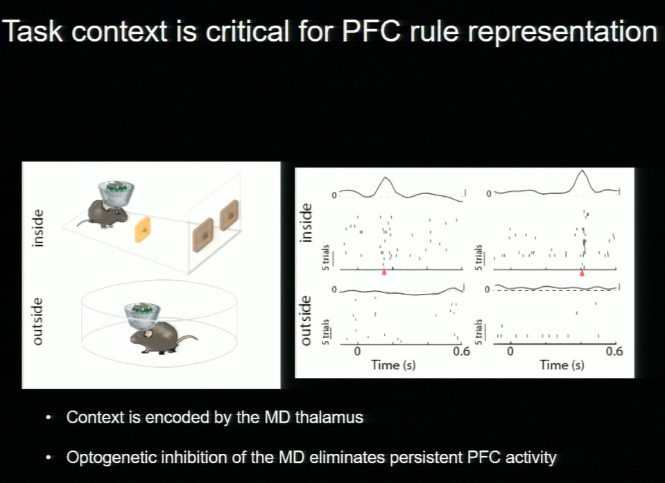
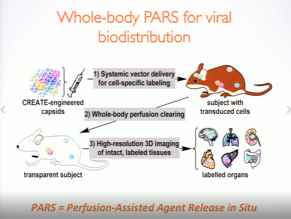
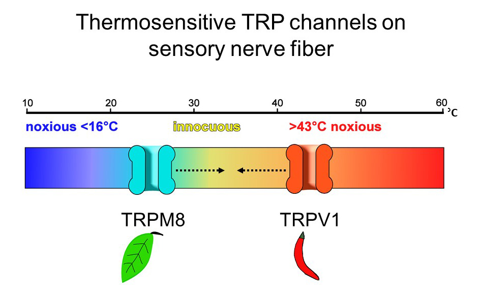
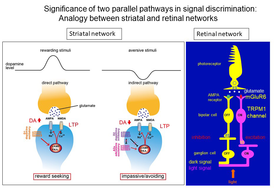
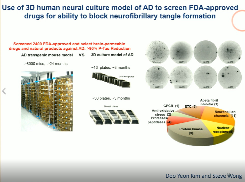
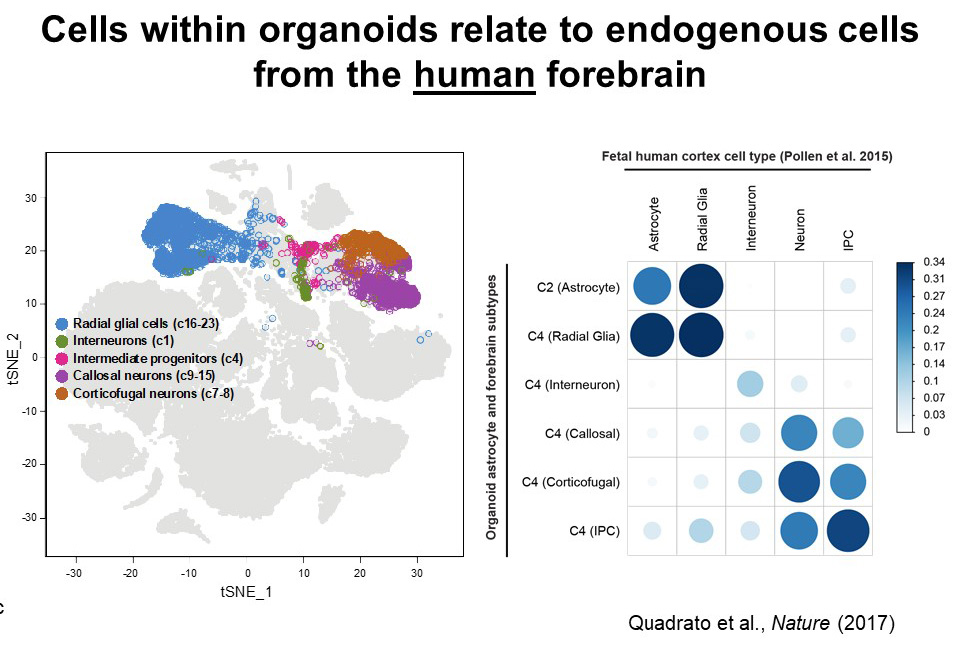
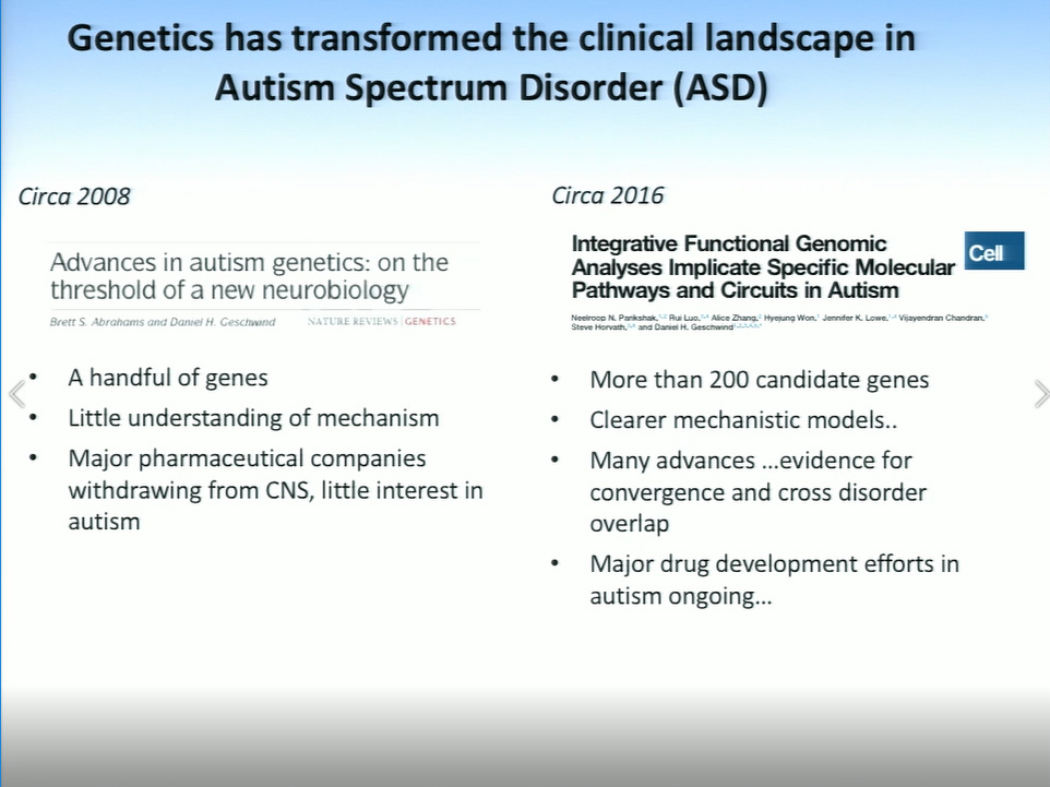


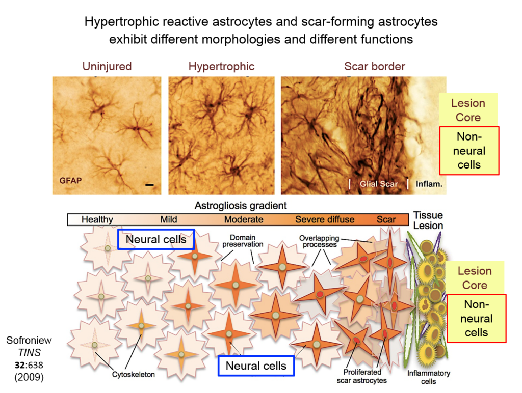

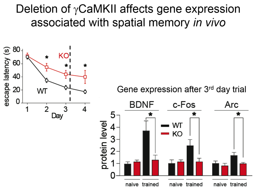
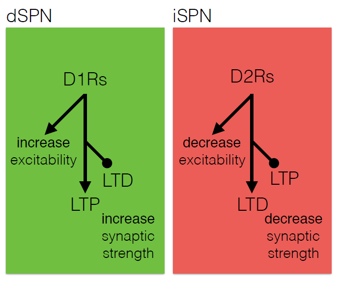
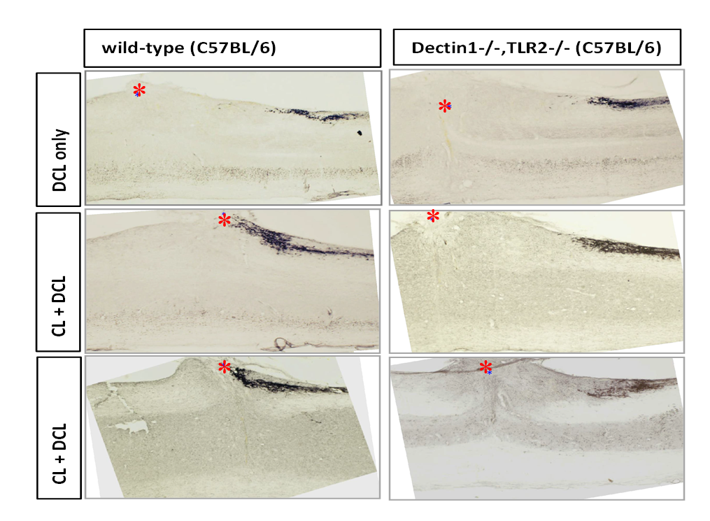
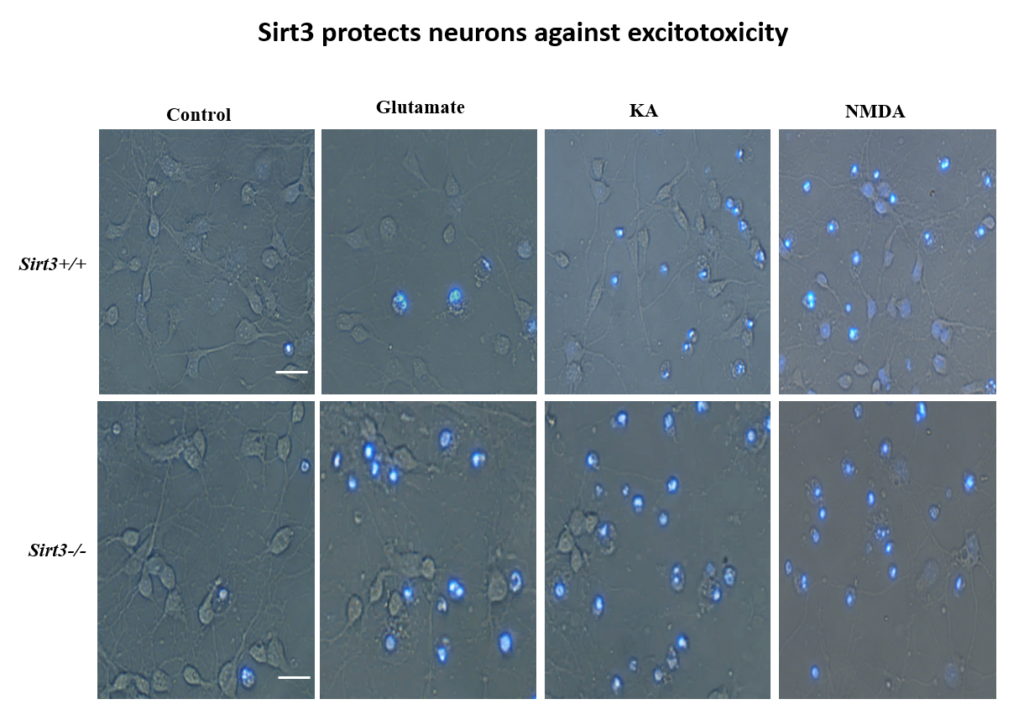
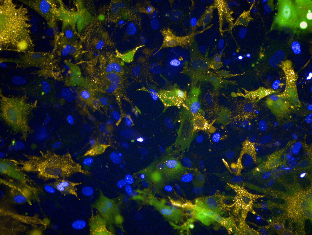
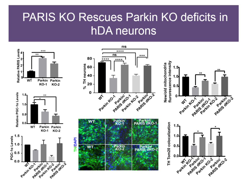
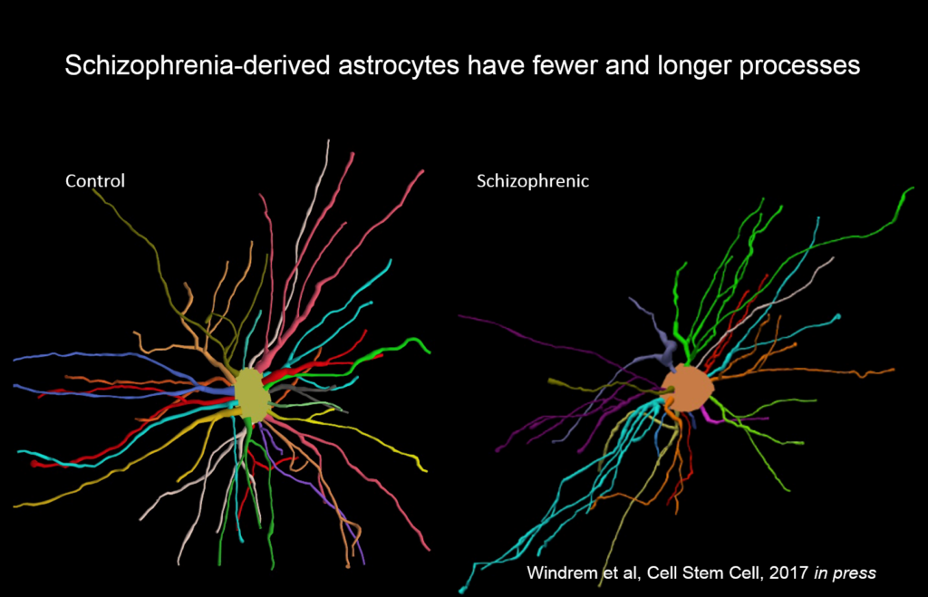
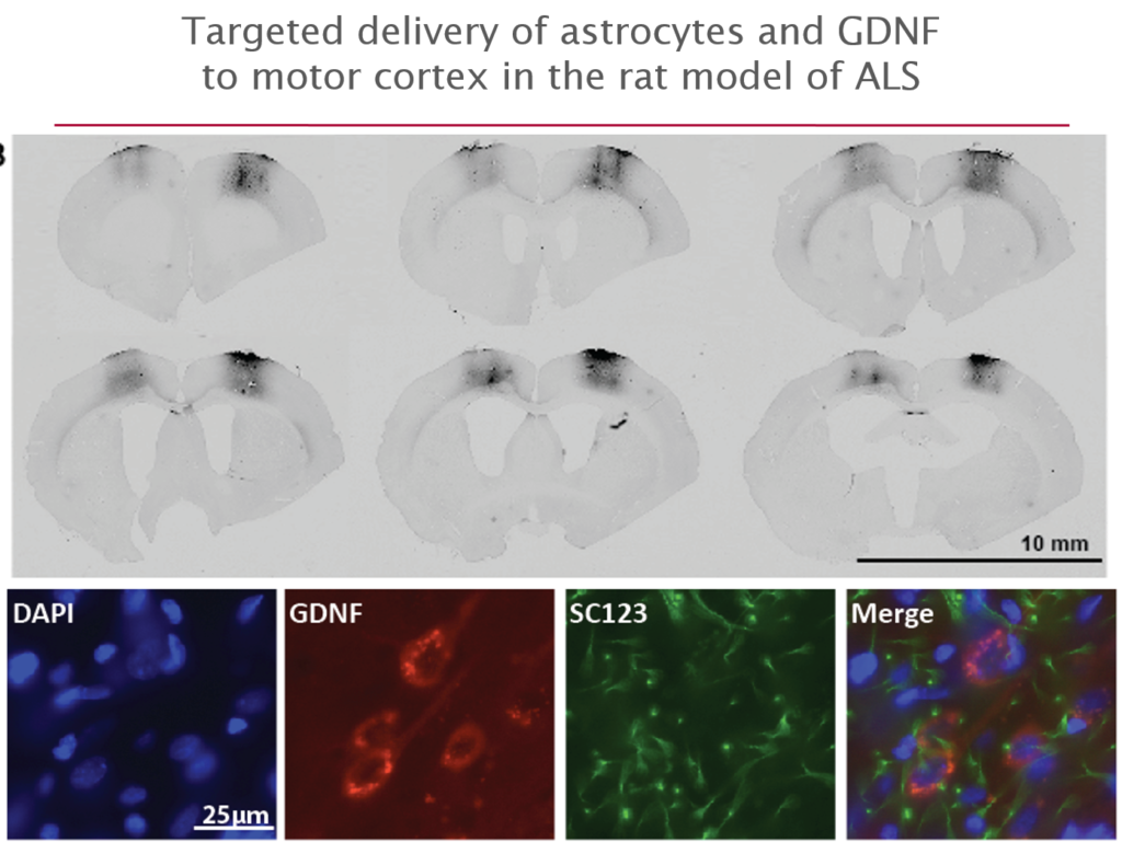
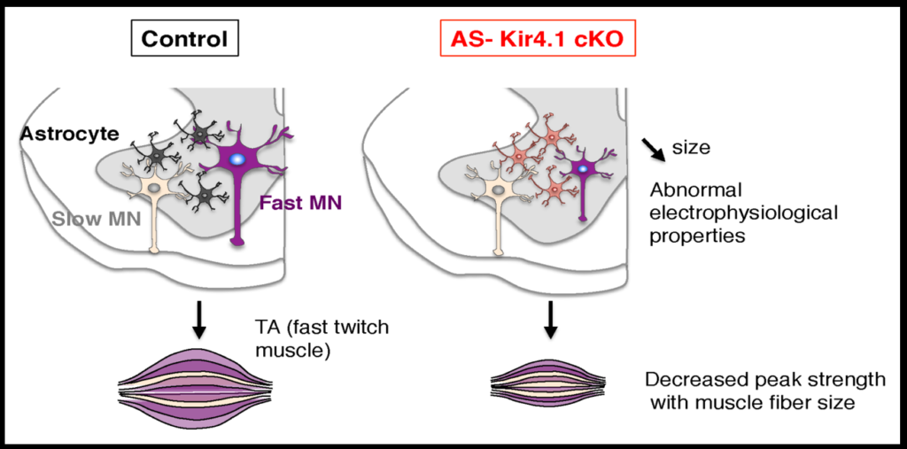
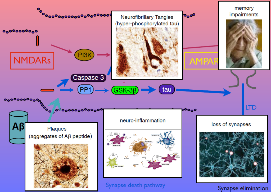
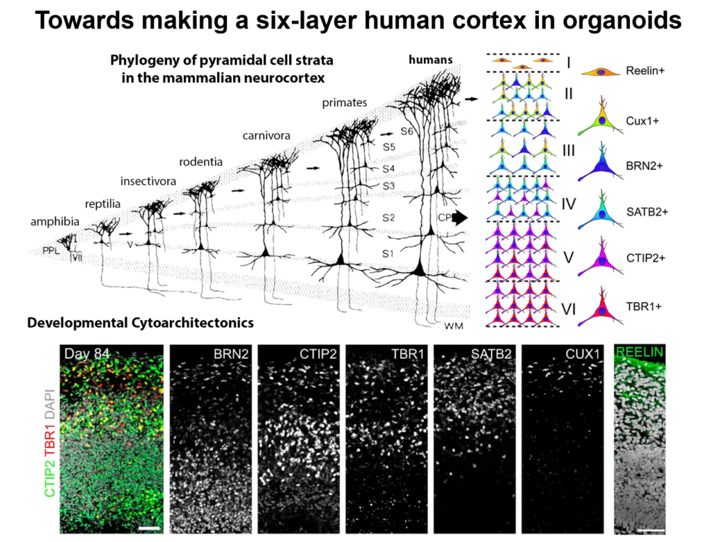
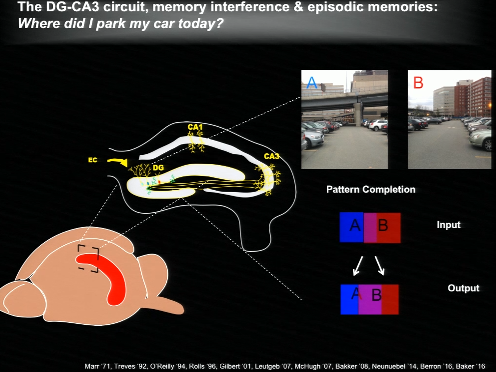
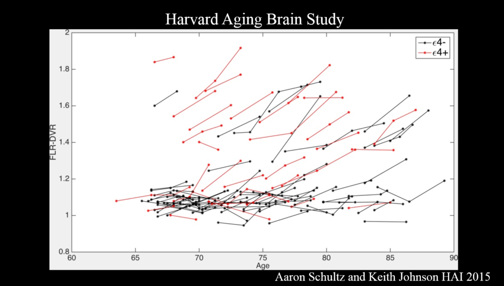
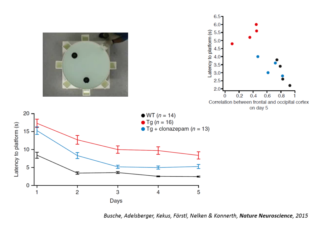
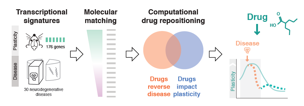


