
Vaccines Strategies for Developing Countries

A mosquito bites a person.

Exploring vaccine treatments for Dengue, Meningococcal, and Pneumococcal, all of which pose a major public challenge in areas like Southeast Asia, Africa, and Latin America.

Published August 11, 2009

By Theresa M. Wizemann

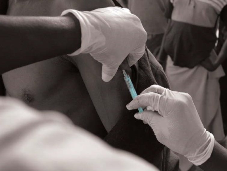
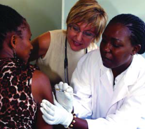
Despite the availability of life-saving vaccines, communicable diseases remain a major public health problem around the world. There is a gap between when vaccines and technologies become available in industrialized countries, and when they are implemented in countries that need them most, where the disease burden is the greatest. This problem persists because of challenges in research and development, prioritization, regulation, funding, infrastructure development, implementation, and surveillance and monitoring.

A May 20, 2009, symposium hosted by The New York Academy of Sciences (the Academy) looked closely at these issues, focusing on meningococcal and pneumococcal diseases, dengue, and dengue hemorrhagic fever. In addition to explaining the state of the art in vaccine development for these diseases, speakers offered several strategies for protecting populations from vaccine-preventable disease: achieve high immunization coverage rapidly and across age groups with the highest disease burden; assess waning immunity; and do not underestimate carriage reservoirs that can reintroduce the organism into the population.

They also discussed the importance of public–private partnerships to ensuring timely, equitable, and sustainable delivery of lifesaving vaccines to developing countries. Some aspects of successful strategies include: regional demand estimates and bulk purchasing, allowing for lowest price negotiation; purchase commitments and annual contracts, allowing manufacturers to plan for production, delivery, and financial investment; and lines of credit to countries, ensuring uninterrupted delivery of vaccines.

Closing the Gap

Despite advances in treatment and prevention, communicable diseases remain a major public health problem in much of the world. Thirty-two percent of all deaths worldwide are due to infectious diseases, and there is a stark inequality in the global disease burden of vaccine-preventable diseases. The developing world, including regions of Southeast Asia, Africa, and Latin America, bear the greatest burden, in part because life-saving vaccines that are part of routine care in industrialized countries still have not been broadly implemented in these regions.

On May 20, 2009, leading scientists from academia, industry, and nonprofit organizations gathered at the Academy to discuss the unique challenges of developing, evaluating, funding, and delivering vaccines for infectious diseases to those that need them most. The symposium, moderated by Albert Ko of Weill-Cornell Medical College, focused on global efforts to prevent meningococcal and pneumococcal diseases, dengue, and dengue hemorrhagic fever.

The challenges of providing these vaccines to the poorest of the world span research and development, prioritization, regulation, funding, infrastructure development, implementation, and surveillance and monitoring. To close the gap, all of these areas must be dealt with concomitantly.

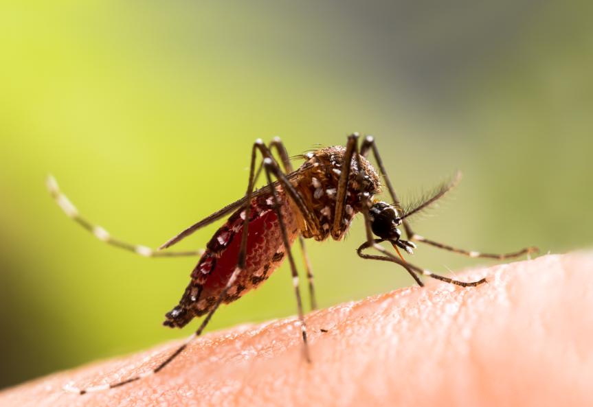
Dengue, Once Nearly Eliminated, Re-emerges with a Vengeance

Dengue hemorrhagic fever is often classified as an emerging infectious disease, Scott Halstead of the Pediatric Dengue Vaccine Initiative (PDVI) said. The often fatal disease has been amplified by factors of the contemporary world. Global population growth, rural-to-urban migration, and deterioration of cities provide breeding space for the dengue vector, the Aedes aegypti mosquito. Jet travel by people unaware they are infected allows the virus to relocate rapidly. In addition, following eradication of A. aegypti from the American tropics in the 1960s in response to yellow fever, the number of vector control experts dwindled and Aedes has reestablished itself in an even wider range than before.

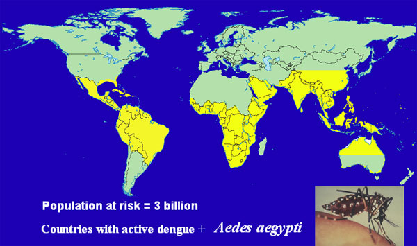
There are four dengue viruses, all endemic in tropical regions of the world. Certain populations, including Africans, appear to have a resistance gene and experience severe disease less frequently. Most observable disease in Africa is in expatriate residents. About half the world’s population, about 3 billion people, live in the tropical regions where the dengue vector thrives. Conservatively, 50–100 million people are infected with dengue each year and about 10% have overt symptoms ranging from mild disease to fatal hemorrhagic fever. The economic impact of dengue illness is considerable.

Countries in yellow are home to the dengue virus, and approximately 3 billion people are at risk.

The majority of severe disease results from secondary infections in those with circulating antibody from a previous infection. Primary infection in infants can also be severe, progressing as if it were a secondary infection due to the presence of maternal antibody. Antibodies interact with dengue virus to form immune complexes that are internalized by monocytic cells, resulting in a complicated phenomenon called intrinsic Antibody Dependent Enhancement (ADE).

In the presence of enhancing antibody (i.e. circulating antibody below protective levels) the number of cells that are infected is greater, and the viral output of each cell is increased. A recent study suggests that while the interferon system is generally effective in controlling infection, in the presence of dengue antibody, immune complexes suppress the interferon system, resulting in more severe disease.

Given the lack of effective vector control and the vast population at risk for contracting dengue, a vaccine is the best hope for controlling the disease. Halstead highlighted five products in development. ChimeriVax-Dengue is a tetravalent dengue vaccine engineered by Sanofi Pasteur by inserting structural genes from the four dengue viruses into an attenuated yellow fever virus. A second tetravalent vaccine being codeveloped by GSK and the Walter Reed Army Institute for Research (WRAIR) is comprised of native whole dengue viruses, attenuated by serial passage.

Two other chimeric tetravalent vaccine candidates are being developed by NIH in collaboration with Johns Hopkins University, and by the CDC in collaboration with Inviragen, based on attenuated dengue 4 and dengue 2 viruses, respectively. Finally, Hawaii Biotech has expressed the four dengue envelope genes in yeast cells. One of the challenges of vaccine development, Halstead said, is targeting the age when maternal antibodies decline sufficiently to allow a live attenuated vaccine to be effective in infants. Achieving herd immunity through mass vaccination is another approach to better protect infants, and to avoid ADE.

The PDVI supports the development of dengue vaccines, preparing field sites for vaccine trials, working to improve and standardize dengue diagnostic tests, and preparing regulatory and manufacturing standards. PDVI is also working to better understand the burden of illness, strengthen surveillance, and upon approval of a vaccine, will advocate for introduction and use, addressing issues of access.

Meningococcus: Preventing Outbreaks and Controlling Endemic Disease

While dengue virus infects only those who come in contact with its mosquito vector, nearly all humans are colonized with Neisseria meningitides at some point. Emil Gotschlich of the Rockefeller University said that about 10% of the population harbors meningococcus asymptomatically in the nasopharynx in winter months, but in schools, prisons, and the military, the carrier rate can be as high as 60%. Since WWII, there has been a relatively low-level endemic pattern of meningococcal meningitis in the U.S., about 2 cases per 100,000 people, with occasional regional outbreaks.

With the essential elimination of Haemophilus influenzae meningitis as a result of vaccination, meningococcus is now the leading cause of bacterial meningitis in U.S. Overseas, epidemic disease is a serious concern. Most prominent is the “meningitis belt” spanning sub-Saharan Africa. There, endemic disease is very common and major epidemics occur at frequent intervals. There is a rapid onset of cases in the winter months, as high as 500/100,000, which halts rapidly when the rainy season starts. This dependence on meteorological conditions is not fully understood.

The meningococcus is covered with a polysaccharide capsule which is required for virulence, and natural immunity is dependent on circulating antibody to the capsule. A relatively limited number of capsular serogroups cause invasive disease, including types A, B, C, Y, W-135, and X. The principle of vaccination with capsular polysaccharide was established by early studies of the pneumococcal vaccine, and trials of first-generation meningococcal polysaccharide vaccines in school-age children and adults established that the efficacy of vaccines to group A and C meningococci was 90%.

However, infants and young children under the age 2, who are at the highest risk of meningococcal disease, do not respond immunologically to polysaccharide antigens. To address this, researchers focused on developing protein conjugate vaccines modeled after the current H. influenzae vaccines. The current meningococcal conjugate vaccine is tetravalent, covering groups A, C, Y, and W-135, and has proven to be highly effective in preventing outbreaks of meningitis, and controlling endemic disease.

Conjugate vaccines are a marked improvement over polysaccharide alone, allowing for vaccination in the first years of life, achieving high levels of antibody at all ages, and effectively suppressing the carrier state. However they do have shortcomings, including the need for boosters to maintain protective levels of antibody. Conjugate vaccines, if properly manufactured, are excellent. What is required, Gotschlich said, is a leadership that devises and promotes programs to match the quality of the vaccine.

The African Meningitis Belt

Despite vaccination in response to epidemics, outbreaks of serogroup A meningococcal disease occur year after year across sub-Saharan Africa, explained Thomas Clark of the Centers for Disease Control and Prevention (CDC). Over 250 million people are at risk across the African meningitis belt, from childhood, well into the fourth decade of life.

The Meningitis Vaccine Project (MVP) was created in 2001 to develop a meningitis vaccine that is effective and affordable for Africa, and ultimately, to eliminate epidemic meningitis as a public health problem in sub-Saharan Africa. A unique consortium of partners is contributing serogroup A polysaccharide, carrier protein, conjugation technology, and manufacturing capabilities. Clinical trials in Africans aged 2–29 years of age have shown that the novel meningococcal A conjugate vaccine, MenAfriVac, is immunologically superior to the existing tetravalent conjugate vaccine, Menactra. Comparable results were observed in children aged 12–23 months.

In the United States, infants under age 1 and adolescents aged 16–19 have the greatest disease burden. Since 2005, CDC has recommended meningitis vaccination at 11–12 years of age, but coverage in adolescents has taken effect more slowly than infant programs, with teen coverage reaching only 30% by 2007.

The U.K. experience is somewhat different. In 1999, in response to the increasing burden of serogroup C disease, the U.K. rapidly implemented immunization of infants, young children, and adolescents, achieving over 80% coverage across all ages by 2001. In 2008, for the first time, there were no deaths from serogroup C meningococcal disease in the U.K. A study of over 10,000 U.K. adolescents showed that vaccination reduced the carriage rate of serogroup C meningococci by two thirds within one year, resulting in herd immunity and a reduction in disease across all age groups, including in those who were not vaccinated.

The Meningitis Vaccine Project’s schedule for implementing mass vaccination across the African meningitis belt.

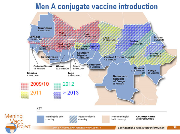
The MVP is modeled on the U.K. experience. Phase 1 is mass vaccination of the population under 30 years of age. Phase 2 includes a catch-up campaign every 5 years in children aged 1 to 5 years, and two infant doses as part of the regular childhood schedule. MenAfriVac has been submitted to the Indian regulatory agency for licensure and a decision is expected in late 2009. Upon approval, Burkina Faso will be the first country to implement vaccination, due in part to the burden of disease, and the ability to conduct surveillance and evaluation. Vaccination will then roll across the meningitis belt.

Even at a cost of 40¢ per dose and about $1 per person to administer, the 12 million doses needed in Burkina Faso are still prohibitively expensive for the country’s ministry of health. Fundraising is ongoing to be able to implement the mass vaccination phase before funding from GAVI is approved. Surveillance will be conducted for coverage levels, vaccine effectiveness, safety, carriage and transmission, outbreaks, and molecular epidemiology, including the emergence of other serogroups.

Pneumococcus: The Number One Cause of Vaccine-preventable Death

Like meningococcus, the primary virulence factor of Streptococcus pneumoniae is the polysaccharide capsule. Pneumococcus is carried asymptomatically in the nasopharynx, and causes clinical disease when it moves to other parts of the body. The most common manifestation is otitis media, infection of the middle ear. More invasive and severe outcomes include pneumonia, bacteremia, and meningitis. Conjugation of pneumococcal polysaccharide to a carrier protein elicits protective levels of functional opsonophagocytic antibody in infants and children.

There are over 90 pneumococcal serotypes, but not all are associated with disease, explained Emilio Emini of Wyeth Pharmaceuticals. The current conjugate vaccine, Prevnar, covers the seven serotypes that are responsible for 80% of invasive disease in children in the U.S.: 4, 9V, 6B, 14,19F, 18C, and 23F. Manufacturing the 7-valent vaccine is extremely complex as it is, in essence, seven separate conjugate vaccines. In clinical trials, the vaccine was shown to be greater than 95% efficacious for invasive disease, and by 5 years after introduction, there was a 98% reduction in invasive pneumococcal disease due to covered serotypes in children. There was also a 76% reduction in disease in unvaccinated adults as a result of herd immunity.

Children in the U.S. have been routinely immunized since 2001; however S. pneumoniae remains the number one cause of vaccine-preventable death in children worldwide. One million infants, and one half million adults, die every year, in large part because the vaccine is just now beginning to be introduced in developing world countries.

The 7-valent vaccine was developed based on serotype prevalence in North America, and the two serotypes that cause epidemic outbreaks of disease in developing world countries, types 1 and 5, are not covered as they are not typically endemic in developed countries. Development of a 9-valent vaccine was undertaken to help address this, and two studies, in South Africa and Gambia, showed the vaccine to be highly effective. The 9-valent vaccine was not fully developed, however, as it became apparent that the epidemiology of the infection was shifting and coverage of more than nine serotypes was needed.

Efforts are now focused on developing a single second-generation vaccine that will be effective worldwide. A new 13-valent vaccine will include serotypes 1 and 5, as well as 3, 6A, 7F, and 19A. All clinical trials have been completed and regulatory approval is now being sought in the U.S. and the EU.

For a vaccine of this complexity, the biggest obstacle to effective delivery to the developing world is manufacturing and cost. The estimated number of pneumococcal vaccine doses required for worldwide use could be as high as 250 million per year. The burden is on biopharmaceutical scientists, Emini said, to improve processes and decrease costs. In addition, WHO requirements must be met before the vaccine is “pre-qualified” (i.e., eligible for purchase by agencies such as the UN that supply vaccines for developing countries). Cold chain requirements also add complexity to delivery and distribution. Emini also noted that political will in the countries in which the vaccine will be made available is required.

In 2009, Wyeth donated enough 7-valent vaccine to immunize every child in Rwanda, which has one of the highest incidences of pneumococcal disease and death. Given the significant unmet medical need, it was decided to begin an immunization program with the current 7-valent vaccine, and transition to the 13-valent as soon as it is licensed.

Breaking the Cycle of High Price, Uncertain Demand, and Limited Supply

Arthur Reingold of the University of Calfornia, Berkeley stressed the dramatic public health impact of the 7-valent pneumococcal vaccine, noting the decrease in disease due to antibiotic-resistant strains, the reduction in noninvasive syndromes such as otitis media, and the decrease in hospitalizations of children due to pneumonia. Children are the primary source of infection for adults, and childhood immunization has led to a significant decline in invasive disease in adults aged 65 and older due to the 7 vaccine serotypes. Remarkably, the indirect herd protection of older children and adults has prevented more disease, and saved more lives, than has direct protection of vaccinated children.

The 7-valent vaccine is administered as three infant doses and a fourth booster dose after age 1 year. But a three-dose schedule could result in a cost savings of 25%, free up global supply, and mean fewer injections for children. Immunogenicity studies have shown that three- and four-dose schedules produce similar antibody levels for most serotypes, and persistence of antibody is similar. The key issue, shown in a case-control study, seems to be that one of the doses must be a booster after the first year of life. Reingold noted that some countries in the EU are already using a three-dose schedule.

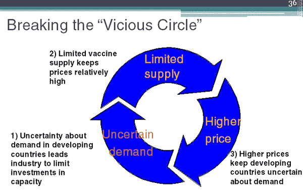
The WHO Strategic Advisory Group of Experts (SAGE) recommended introduction of the 7-valent vaccine in the developing world now, switching to the 13-valent when licensed. The challenge now is implementation. There is, in essence, a vicious cycle of high price, uncertain demand, and limited supply, Reingold said.

A cycle of structural pressures makes it difficult to get vaccines to where they are needed most.

PneumoADIP is working to break this cycle through the development of public–private partnerships. Eligible countries, those with annual per capita income levels of less than $1000, first commit to using the vaccine. Manufacturers, then assured of a market, commit to making and selling the vaccine at a lower price. GAVI and other funders commit to purchasing the vaccine at the guaranteed price for the countries, with a minimal per dose copayment from the country.

This approach, while assisting high-burden countries, does not assist mid-income level countries in the Americas that are not GAVI-eligible, yet cannot afford to pay full price for the vaccine doses they need to give. Some are considering cutting costs by administering fewer doses per child, the health impact of which remains to be seen.

Funding and Delivery: The High Cost of Inaction

Describing his work at the Pan American Health Organization (PAHO), Jon Andrus began by noting that the introduction of vaccines in the Americas (Latin America and the Caribbean) has eliminated or eradicated measles, neonatal tetanus, and polio, and significantly reduced the incidence of diphtheria and pertussis. There is still work to be done, however. In the Americas, two children die every hour of pneumococcal invasive disease. The 7-valent vaccine could prevent at least one of those deaths every hour, Andrus said.

The PAHO experience shows that new vaccines can be successfully introduced and sustained over time. The PAHO Revolving Fund was established 30 years ago to ensure a safe, effective, uninterrupted, sustained supply of vaccines. The Fund uses a bulk purchasing strategy, negotiating best price based on regional demand, rather than per country estimates. A credit system allows for delivery of vaccines in the absence of immediate funds. Countries reimburse PAHO within 60 days after vaccine is delivered, and pay 3% of the price of the vaccine as a service charge to be put into PAHO’s working capital.

This allows for stable and prompt supplies of safe, high-quality, affordable vaccines. For manufacturers, annual contracts facilitate the ability to plan for production, delivery and financial investment. There are reliable demand forecasts, and transparency of relationships. PAHO facilitates “one-stop shopping” for manufacturers, eliminating the need to negotiate individually with 36 member states.

In 2006, the Directing Council of PAHO passed a landmark resolution urging all member states to identify new revenue sources to sustainably finance the introduction of new vaccines against rotavirus, pneumococcus, influenza, and human papilloma virus (HPV). The Revolving Fund policy of single best market price, linked with the power of bulk purchasing, will be critical to accelerated and sustained uptake of these vaccines in participating Latin American countries, Andrus concluded.

If the introduction of pneumococcal and rotavirus vaccines is delayed, almost one million children could die from vaccine-preventable diseases over the next 30 years, and the world would not achieve MDG4, the Millennium Development Goal to reduce child mortality. Ensuring equitable and sustainable introduction of new vaccines into developing countries requires a strategic vision grounded in long-term goals, Andrus emphasized, not short-term fixes.

Moving forward

In summary, to successfully protect populations from vaccine-preventable disease, initiatives should strive to achieve high immunization coverage rapidly, across age groups with the highest disease burden, and should monitor waning immunity. The impact of herd immunity in controlling infectious diseases can be considerable. (i.e. the indirect protection of unvaccinated individuals in a population where a sufficiently high percentage has been vaccinated). However, carriage reservoirs that can reintroduce the organism into the population should not be underestimated. Working as partners, the public and private sectors must develop creative funding approaches to ensure timely, equitable, and sustainable delivery of lifesaving vaccines to developing countries.

Also read: Maternal, Neonatal, and Early Infancy Vaccine Developments

Helping Physicians Better Understand Genomics

A DNA helix.

A new initiative will include an array of efforts, such as a series of ongoing symposia and online community-building tools.

Published June 18, 2009

By Adrienne J. Burke

Image courtesy of Anusorn via stock.adobe.com.

The Life Technologies Foundation has awarded The New York Academy of Sciences (the Academy) a two-year, $400,000 grant to help educate thousands of physicians and medical students in how to utilize new technologies and share cutting-edge research to improve healthcare and understand disease.

“There is a critical need to educate our physicians, both the current generation and the next, to use genomics to think about disease and treatments, and to apply the latest discoveries in this field to the understanding of human health,” said the Academy’s Scientific Director Stacie Grossman Bloom.

To do this, the Academy will build a live and virtual, global community of physicians, medical students and scientists, focused on the pressing issues and challenges of using new technologies in medicine. Organizers hope this effort can serve as a model of improving physician education and health outcomes in urban centers throughout the nation.

The grant was among three that the Foundation made to organizations working to advance science. “We are proud to support these innovative organizations that advance scientific understanding,” said Greg Lucier, Chairman and CEO of Life Technologies. “The grantees we have chosen are working to demystify science to the public by providing training and access for using biology to make life even better.”

The Life Technologies Foundation is a non-profit arm of Life Technologies Corporation, a global biotechnology tools company dedicated to improving the human condition.

Also read: Collaboration Is Key to Breaking New Ground in Genomics

A Global Giver Lends Support from Japan

A shot of beautiful architecture and cherry blossoms in Japan.

With a successful medical career in obstetrics and gynecology, Kenichi Furuya also spends his time advancing science as a member of the Academy’s Darwin Society.

Published March 1, 2009

By Adelle C. Pelekanos

Image courtesy of ake1150 via stock.adobe.com.

At the core of The New York Academy of Sciences’ (the Academy’s) mission is a commitment to “creating a global community of science for the benefit of humanity.” It is a statement that deeply resonates with the Academy members from 140 countries – including Darwin Society member Kenichi Furuya. For this Japanese researcher, the Academy membership is one important way to bridge the distance between Tokyo, New York City, and other international hubs of science.

Furuya, a specialist in obstetrics and gynecology, holds both an MD and PhD. He is a professor and Chairman of the Department of Obstetrics and Gynecology at Japan’s National Defense Medical College. In addition to his association with the Academy, Furuya is a fellow of the International College of Surgeons (headquartered in Chicago) as well as a number of Japanese medical societies. He was born in Tokyo in 1953, and still lives in a central area of the city, Bunkyoku.

A Proud Scientific Tradition

Furuya graduated from the School of Medicine at Japan’s Juntendo University in 1979. He recounts his alma mater’s history with pride: “Our medical school was founded as one of the oldest western-style private hospital/schools in Edo City (Tokyo), in 1838,” during a period of national isolation. Thirty years later, Japan’s Meiji Revolution opened the country’s doors to the West, Furuya explains. Juntendo’s third president, Susumu Sato, was the first Japanese student to study abroad officially, and since the late 19th century the school has encouraged international education and collaboration between researchers. Furuya is a product of this tradition, as evidenced by his active membership and generous support of the Academy.

In the almost 30 years since graduating from Juntendo, Furuya has worked in various areas within obstetrics and gynecology, including basic molecular research, reproductive immunology, clinical reproductive medicine (such as IVF- ET and laparoscopic surgery), and clinical pelvic surgery (such as ovarian and uterine cancers).

In his current work, Furuya focuses on two areas of gynecological research. First, he is studying the mechanisms by which the fetal period of pregnancy (week 10 through birth) affects the development of metabolic disorders in children. In particular, Furuya is interested in diabetes, obesity, and hypertension as epigenetic influences of this period, in pregnancies complicated by placental malfunctions such as gestational diabetes mellitus, nutritional deficiency, and pregnancy-induced hypertension.

Secondly, Furuya is working to clarify the basic mechanism of the relationship between ovarian endometriosis (EM) and ovarian cancer. Epidemiologic findings indicate a strong positive correlation between ovarian EM and ovarian clear cell carcinoma characterized as “refractory,” or resistant to chemotherapy, he explains.

It Runs in the Family

Kenichi Furuya

Furuya’s family, past and present, shares the doctor’s dedication to medicine. Furuya’s wife is an anesthesiologist, his son is an obstetrics-gynecology resident, and his daughter is in dental school. His late father, Hiroshi Furuya, was a gynecologist and emeritus president of the Society of Tokyo Maternal Health. In the 1970s, the elder Furuya was a visiting professor at Germany’s Hamburg University, as well as Columbia University. Furuya not only inherited his father’s vocation, but also his passion for participation in the global science community. It was his father’s status as an Academy member during his time at Columbia that inspired Furuya to become a member 20 years later.

Support in an Important Time

Furuya’s proud support of the Academy conveys his passionate support for scientific collaborations across the globe, and in particular, between the US and Japan. With the new presidential administration, Furuya believes that the American society may be undergoing its “fourth revolution”—identifying the first as the American Revolution, the second as the Civil War, and the third as the end of World War II. “I have been impressed indeed that [the US is changing its] basic social, political, and historical foundations,” Furuya explains. He likens this period in American history to his own country’s Meiji Revolution, the time that ushered in new world views and sparked international dialogue between Japan and the world. Furuya’s long-distance membership is his vote of confidence in the current and future relationship between the US and Japan.

Although Furuya has traveled to New York a number of times, he has not been to the new Academy headquarters at 7 World Trade Center. He plans to visit in the near future, and to continue his support of the Academy. “It is my great honor to support the activities of The New York Academy of Sciences given its long history and many pure science traditions,” Furuya says.

Also read: Changing the Face of Molecular Medicine


About the Author

Adelle C. Pelekanos is a freelance science writer in New York City.

A New Approach to the Hippocratic Oath

A variety of different pills and other medicine in over-the-counter packaging.

For more than 40 years, public- and private-sector biochemical pharmacology experts have been sharing knowledge at Academy meetings.

Published September 1, 2007

By Jill Pope

Image courtesy of Artinun via stock.adobe.com.

It’s a rare occasion when scientists from competing pharmaceutical companies and academic laboratories come together to share their latest findings on human diseases and treatments. But since 1964, The New York Academy of Sciences (the Academy) has played host to a regular meeting of biochemists, molecular biologists, and biomedical researchers who do just that.

The members of the Biochemical Pharmacology Discussion Group (BPDG) hail from more than a dozen pharma and biotech companies, as well as top research universities and medical centers. Pfizer and Bristol-Myers Squibb provide major funding. The American Chemical Society, AstraZeneca, Boehringer Ingelheim, and Novartis also sponsor the group.

As the oldest of the Academy’s 14 discussion groups, the BPDG convenes eight times a year for half- and full-day symposia where experts address topics designated by the group’s steering committee. More than 70 scientists attend each meeting.

Good Career Move

Academy Fellow Martha Matteo began attending the group’s meetings nearly 25 years ago when she was a scientist for Boehringer Ingelheim. New to the pharma industry in 1983, she recalls she was pursuing a theory about how anti-inflammatory steroids affect protease levels. It ran counter to the conventional wisdom that “leukotrienes and prostaglandins modulate everything.”

She organized a BPDG meeting where other scientists presented evidence that steroids induce protease inhibitors. “The story was just unfolding and I got right in the thick of it,” she says. “I had the opportunity to test ideas with a broad range of industry and academic scientists, separate from long-held beliefs and prejudices.” Matteo, who eventually became director of knowledge management and R&D planning at Boehringer Ingelheim before retiring last year, adds, “The Academy has long provided neutral territory and instant feedback in the exploration of new ideas.”

For industry and academic researchers who work mostly in isolation from one another, BPDG events are opportunities to connect, says Charles Lunn, a research fellow at Schering-Plough Research Institute and the group’s current program coordinator. While attendees don’t disclose proprietary information, people do share their work. That’s essential because in industry, Lunn says, “Much high-quality science is accomplished that is never communicated to the academic community.”

From Theory to Therapy

An Alzheimer’s seminar drew more than 100 participants last December. This is another example of a BPDG forum where researchers discuss cutting-edge research. Alzheimer’s researchers have focused on two main culprits in their search for the cause of this devastating disease: amyloid-β peptide (A-beta), which forms plaques in the brain, and tau, a rogue protein that forms tangles. A-beta is produced when a large protein is cut by two enzymes. Several leading experts on one of those enzymes, γ-secretase, shared their insights into how it might be targeted by Alzheimer’s therapies. Others discussed the role of tau: some showed how amyloid pathology may trigger changes in tau, and others examined how tau abnormalities lead to cell death.

Speakers included Mark Shearman, senior director of neuroscience drug discovery research at Merck in Boston; Thomas Lanz, a scientist in central nervous system biology at Pfizer Global Research & Development; Michael Wolfe, who in 2006 established the Laboratory for Experimental Alzheimer Drugs at Harvard Medical School; and David Holtzman, head of the Department of Neurology and associate director of the Alzheimer’s Disease Research Center at Washington University in St. Louis.

Matteo, who chaired the group from 1989 to 1994, says it’s not unusual to see theories presented at BPDG meetings turn into therapies years later. Around 1990, she remembers, the group held a meeting to discuss a potential approach to cardiovascular disease called angiotensin II receptor blockers. Today, ARBs such as losartan and valsartan are standard therapy for hypertension.

Setting The Agenda, Seeking Diversity

It’s easy to imagine how the BPDG will continue to benefit young scientists’ careers the way it did Martha Matteo’s.

Recent seminars have included another on Alzheimer’s research trends—“Immunotherapy for Neurodegenerative Diseases,” in which experts discussed ways to train the body’s immune response to attack the wayward proteins that plague patients with Alzheimer’s and other diseases of the brain and spinal cord.

In May 2007, the group hosted “The Future of Monoclonal Antibody Biotherapeutics.” Monoclonal antibodies are cloned proteins that modulate the activity of specific disease targets. In cancer treatment, they zero in on tumor promoters, leaving healthy tissue alone. The therapies are already benefiting patients, but they have limitations, including high production costs. Speakers discussed new approaches, such as optimizing cell culture processes, that promise to spur the therapies forward and make them more widely available. Also this past year, speakers at BPDG’s “Novel Strategies for Compound Identification from Compound Libraries: High-Throughput Screening” presented diverse approaches to drug screening such as Biotrove’s RapidFire mass spectrometry, and virtual screening with the University of New Mexico’s high-throughput flow cytometry platform.

Diabetes to Stem Cells

The 2007-2008 meeting schedule will cover progress in treating diabetes and eating disorders, psychiatric illness, and atherosclerosis, as well as tools for drug discovery including adult stem cells and molecular imaging. Setting the year’s agenda is a labor-intensive process, requiring committee chairs to tally the votes of hundreds of discussion group members. But the result is worthwhile, says Ross Tracey, an associate research fellow at Pfizer who led the group from 2002 to 2006: “The programs that emerge have clearly passed the popularity and interest test.”

To ensure the continued relevance of BPDG meetings, Jose Perez, a senior principal scientist at Pfizer and a committee co-chair, is on a mission to recruit new members to the group. In the coming year, he’ll reach out to scientists at underrepresented pharma and biotech firms, as well as at New York City’s universities. “That’s the only way the organization is going to have a broad perspective,” he says. “We really strive for diversity of thought.”

Also read: Equivalence of Complex Drug Products: Scientific and Regulatory Challenges


About the Author

Jill Pope writes about science and policy issues. She served as Senior Editor for The Cutting Edge: An Encyclopedia of Advanced Technologies (Oxford University Press, 2000).

How Can Science Help in the Fight Against Poverty?

A straw hut.

A global scientific publishing initiative follows the philosophy of the Millennium Development Goals by tackling poverty from all angles

Published September 1, 2007

By Leslie Taylor

For the last decade, a technological marvel, has been saving lives in sub-Saharan Africa. It has no bells and whistles, no microprocessors or moving parts. It is a simple piece of insecticide-treated netting.

Bed nets made from this material remain effective deterrents against mosquitoes for three to five years. Donors, governments, and community leaders have embraced the low-tech tool as a valuable public health intervention and frequently hand out nets during immunization campaigns and antenatal clinics. About $5 buys a net that will shield two children from mosquitoes as they sleep—an incredibly effective means of preventing malaria, a disease that kills more than 1 million people a year.

The nets are a great example of what can be achieved when the scientific and development communities work together to identify needs and implement new ideas, says John McArthur, who was deputy director of the United Nations Millennium Project and is now associate director of the Center for Globalization and Sustainable Development at Columbia University’s Earth Institute. To put life-saving technology in the hands of the people it is designed to benefit requires the cooperative efforts of scientists, policy makers, and the communities they hope to serve, he says.

A Different Publish-Perish Paradigm

That philosophy of partnership underpins the Millennium Development Goals, which aim to achieve target levels of world-wide nutrition, health, literacy, and environmental sustainability that were set at the Millennium Summit in September 2000. It is also at the heart of a new program called Scientists Without Borders SM that was co-conceived by The New York Academy of Sciences (the Academy) and the U.N. Millennium Project. And now a massive cooperative effort in the interest of global development is taking place among scientific publishers.

This year, halfway to the 2015 deadline that world leaders set for achieving the Millennium Development Goals, 230 science journals worldwide will simultaneously publish papers or special editions on the topic of poverty and human development. Publications participating in the Council of Science Editors initiative include wide-circulation journals such as Science and Nature and more specialized volumes such as the African Journal of Drug and Alcohol Studies, the Chinese Journal of Evidence-Based Medicine, and the Wisconsin Medical Journal.

The Annals of the New York Academy of Sciences will publish a volume titled Reducing the Impact of Poverty on Health and Human Development: Scientific Approaches.

A Multidisciplinary Approach

The Annals volume, edited by Stephen Kaler and Owen Rennert of the National Institute of Child Health and Human Development, takes a multidisciplinary look at the issues facing the world’s poor. Chapters address public health issues in the developing world as well as specific diseases associated with poverty, such as tuberculosis, malaria, HIV/AIDS, lymphatic filiariasis, and hookworm. Other chapters discuss the poor’s access to health care services, education, proper nutrition, and housing.

The volume will highlight diverse areas of research. It will include a paper on measles by Samuel L. Katz, chairman emeritus of pediatrics at Duke University, who was awarded the 2007 Pollin Prize in recognition of his contributions to pediatric infectious disease research and vaccine development; a paper titled “Sustainable Transfer of Biotechnology to Developing Countries,by Eva Harris, who used the money from her 1997 MacArthur “Genius” Fellowship to establish the Sustainable Sciences Institute, an organization that helps scientists around the world gain access to state-of-the-art training and equipment; and a paper by Nobel Laureate James J. Heckman, professor of economics at The University of Chicago, about the consequences of poverty for human skill formation.

Poverty Is a Many-Stranded Problem

Bashir Jama, author of “Agriculture in Developing Nations,” a paper in the upcoming Annals volume, spent 19 years with the International Centre for Research in Agroforestry before becoming a policy advisor to a U.N. Development Program group working on poverty and the Millennium Goals. He says it’s very difficult to tease apart the problems of poverty and address any single factor in isolation. Agriculture is inextricably linked to health, he says.

For instance, malaria and other tropical diseases can impede worker productivity in farming communities, resulting in reduced crop yields, followed by hunger, and increased vulnerability to disease.

And illiteracy can be an obstacle to heartier harvests. Training in new farm techniques or agriculture technologies can’t be distributed in writing to farmers who can’t read, he notes. Instead, non-governmental organizations and governments must offer in-person training or demonstration farms.

“As scientists we have fairly good knowledge of the ecology and the technical issues that are slowing down progress or that can enhance production,” says Jama. “But giving people the skills they need when they live in remote areas—in areas with limited energy supplies, no electricity or clean water—is challenging.”

Within select communities known as Millennium Villages, networks of scientists with diverse areas of expertise work with residents to address the intertwining issues of agricultural productivity, health, education, and access to markets. Projects to increase food yields and improve access to education and health services coincide with initiatives to improve village infrastructure—roads, sanitation, communication technology, and energy. Villagers are also given advice on enterprise diversification and environmental management.

Leverage Existing Technologies

Residents of the 12 Millennium Villages in 10 African countries have seen tremendous improvements in quality of life since the project started, Jama says. “In one or two growing seasons we’ve seen incredible increases in agricultural productivity, phenomenal decreases in hunger, improved health with a reduction in malaria and waterborne diseases, and safe drinking water becoming available,” he says.

Successes at the Millennium Village sites were not the result of exclusive breakthrough technologies, but came about because experts in a variety of fields took action to supply villagers with a range of basic technologies, such as fertilizer, medication, and water purification systems. “We have the basic know-how,” says John McArthur. “The question in the immediate term is how to mobilize existing technologies.”

Frequently, technologies created for another purpose or discovered in the course of pure research can be greatly beneficial. “It’s a matter of adapting good technologies that may exist in other countries,” says Bashir Jama.

Seemingly uncomplicated technology can have a dramatic impact. For example, the treadle pump—an inexpensive, simple- to-operate, foot-powered pump that can draw water from a well or spring—has revolutionized farmers’ ability to grow food during the dry season. “It’s a good example of a situation where, if the investment is there, it could really increase irrigation, and improve income and nutrition,” says Jama.

Energy and Resource Use

Improved cook stove technologies have also done much to improve the lives of the poor, according to Daniel Kammen, a professor in the Energy and Resources Group at University of California, Berkeley, who contributed a paper titled “Energy & Resource Use in Developing Countries” to the new Annals volume. Respiratory illnesses are one of the biggest health problems in the developing world, where most people typically cook using very simple fires—burning wood or dung on just a few stones. “Making stoves more efficient has actually cut down on one of the leading causes of illnesses worldwide,” he says.

Kammen, who is also founding director of the Renewable and Appropriate Energy Laboratory, an organization that focuses on designing, testing, and disseminating renewable and appropriate energy systems, has seen how the timely application of technology can transform communities. His group works on projects such as promoting sustainable biomass energy management in Zimbabwe, evaluating the performance of single junction amorphous silicon modules used in photovoltaic systems in Kenya, and creating new technologies such as the UV-Tube—an inexpensive and easy-to-use household water disinfection device that uses ultraviolet light to inactivate pathogens.

While each country has slightly different needs, Kammen explains, in most parts of the developing world the basic issues are the same. “There’s a lack of access to clean water, a lack of electricity to do things like read at night or run a business, and a lack of access to education,” he says. “There are some constants, and those mean you can work pretty hard on a project in one country and it’s likely to be useful to people in many other parts of the world. It’s not like a solution you develop in Mozambique is only useful there.”

Create New Technologies

For problems of the poor that do not yet have technological solutions, scientists have found new ways to obtain funding to do the research they hope will ultimately alleviate suffering.

Peter Hotez, editor-in-chief of a soon-to-launch Public Library of Science journal called Neglected Tropical Diseases, wrote a paper about hookworm for the Annals volume. He is president of the Sabin Vaccine Institute, a nonprofit organization that works to provide the world’s poorest people with access to low-cost, safe vaccines and drug treatments for neglected tropical diseases—13 parasitic and bacterial infections that produce chronic and disabling conditions. Many people have not heard of the diseases—including scariasis, hookworm infection, trichuriasis, lymphatic filariasis, onchocerciasis, schistosomiasis, and trachoma—but they are devastating.

“Neglected tropical diseases are one of the primary reasons why poor people remain poor. In some ways [what they do to a person] is worse than death,” says Hotez. “They destroy quality of life and are one of the major reasons we have poor economic development in Africa and elsewhere. These are the diseases that are keeping people mired in this horrible cycle of destitution and despair.”

Yet, until recently, little attention was paid to these scourges. While the private sector has been willing to invest money in research that might lead to an AIDS vaccine, for which there is still a substantial market in the U.S. and Europe, “There’s no way you could ever make a profit on a hookworm vaccine,” says Hotez.

Vaccines and Medication

Thankfully, the Human Hookworm Vaccine Initiative, a public development partnership sponsored by the Sabin Vaccine Institute with major funding from the Bill & Melinda Gates Foundation, is working to develop and disseminate an effective, safe, and low-cost vaccine. “It’s a unique model for making a product for people who can’t afford to pay for it,” Hotez says.

While the vaccine is not yet ready to be distributed, the Global Network for Tropical Disease Control, a program of the Sabin Institute, distributes a “rapid impact” package of medication that includes four anti-parasitic drugs to treat seven neglected diseases. The health kit, which costs only 50 cents per person per year, greatly reduces rates of morbidity, blindness, and skin disease. Yet it is only a short-term solution because diseases such as hookworm have high rates of transmission and re-infection, Hotez explains.

“Millennium development goal number six is ‘to control and fight HIV/AIDS, malaria, and other diseases.’ We feel we can make an impact right now in the ‘other diseases’ category,” he says.

Questions of Investment: Time and Money

While sufficient will and technologies are available to raise the standard of living in the developing world, funding is a primary barrier to success. Too little money is devoted to the cause, and there is no consensus about how the money that is devoted should be spent, experts say.

“A rule of thumb, which varies a little by country and by need, is that it takes a basic investment of about $110 per person per year to achieve the goals outlined in the Millennium Development Project,” says John McArthur. “Right now there is, on average, $25 per person in foreign aid going into these places. That needs to be scaled up two- or three-fold by 2015. There’s not enough money getting to where it needs to go, and a greater share needs to go to practical technologies, like long lasting insecticide-treated bed nets, fertilizer, or drilling bore wells.”

The Need for Collaboration

Bashir Jama worries that, too frequently, what scientists have discovered about issues of development is not being incorporated into national, regional, and global programs. “Decisions are made in a vacuum as though science doesn’t exist,” he says. “Donors, international governments, the policy makers need to take advantage of this knowledge and to link up better with scientists in designing systems that work.”

At the same time, it is important for scientists to make the effort to collaborate with policy makers and with one another in the fight against poverty, suggests Hotez, sharing this quote from Dr. Albert Sabin, the inventor of the polio vaccine, after whom the Sabin Institute is named: “A scientist who is also a human being cannot rest while knowledge which might reduce suffering rests on the shelf.”

Also read: Scientists Step into New Roles to End Poverty


About the Author

Leslie Taylor is associate editor of Update and of the Academy’s online public gateway, Science & the City.

Organic Morality: Our Intuitive Inheritance

That graphic in the shape of a human brain.

In a new book, the Harvard evolutionary psychologist argues that all humans share an innate sense of right and wrong.

Published March 1, 2007

By Laura Buchholz

You are in control of a switch at a railroad station. An empty out-of-control train is racing toward five people walking on the tracks. It will hit and kill them unless you pull a lever to switch the train to another track—but there it will kill one person standing on the track. Do you pull the lever? Why? Or why not?

You are an emergency room doctor. Five of your patients urgently need organ transplants in order to live. In the waiting room is a healthy young man with all of the organs necessary to save these five people. Would you sacrifice the life of the man to save your five patients? Why? Or why not?

If you answered “yes” in one case and “no” in the other, what is the difference between the two cases?

Marc D. Hauser, professor of psychology, organismic and evolutionary biology, and biological anthropology at Harvard University, explores how we answer questions like these in his new book, Moral Minds: How Nature Designed Our Universal Sense of Right and Wrong. On January 11, 2007, as part of The New York Academy of Sciences’ (the Academy’s) Readers & Writers series, Hauser explained that moral decision-making may not flow entirely from experience and education, but instead may have a significant biological aspect that has been shaped, like all human traits, by the forces of evolution.

Instinctive Morality

“We are endowed with a moral faculty evolved to generate intuitive judgments of right and wrong,” says Hauser, adding that the principles underlying those intuitive judgments are unconscious, and therefore, may be immune to cultural influence. In other words, Hauser suggests that the influence of Sunday school may pale in comparison to the effect of thousands of years of genetic programming.

Hauser, who directs the Cognitive Evolution Laboratory at Harvard, collects some of his evidence from the Moral Sense Test—a Web site his lab developed, presenting visitors with “artificial dilemmas” designed to test their moral instincts. Working with a data set of responses from 250,000 subjects from 120 different countries, with ages ranging between 13 and 70, and inclusive of all varieties of religion, Hauser’s lab finds some patterns emerging.

Hauser identifies three principles of automatic moral reasoning that transcend religion, geography, age, and culture. The first is the Intention Principle: That is, most people judge it morally worse when harm is intended as a means to an end as compared with when an equivalent harm is foreseen but as a side effect. When Joe intentionally hits John we tend to hold him more responsible than when Joe strikes an object with the foreseen consequence that this object will fall and hit John. According to Hauser, the Intention Principle operates at an unconscious level: When people judge based on this principle, they are not able to say why they made the judgment.

The second principle Hauser calls the Action Principle, and it states that harm caused by action is worse than exactly the same harm caused by omission.

Consider Two Scenarios:

#1: A man intends to kill his young nephew, who stands to inherit all the family wealth. The uncle goes up to the bathroom where the boy is taking a bath, and drowns the boy in the tub.

#2: A man intends to kill his young nephew, who stands to inherit all the family wealth. The uncle goes up to the bathroom where the boy is taking a bath, and finds the nephew drowning face-down in the tub. The man does not intervene, and lets the boy drown.

The effect is the same, but would a jury find the uncle guilty of murder in the second scenario? Probably not. This principle is available at a conscious level, says Hauser, and may explain why societies generally find active euthanasia more morally troubling than passive euthanasia.

Third is the Contact Principle, which states that harm caused by contact is morally worse than equivalent harm caused by non-contact (e.g., when we hit someone vs. seeing an object fly across a room and hit somebody—or the difference between the two introductory scenarios). This third principle is partially available to human consciousness—about half and half, says Hauser.

Remarkably, Hauser notes that subjects who described themselves as highly religious delivered the same judgments as those who said they were not at all religious. These observations suggest that the system that unconsciously generates moral judgments is immune to religious doctrine. But what does this have to do with biology? Hauser draws a parallel between what he calls our “universal moral grammar” and Noam Chomsky’s linguistic theory of universal grammar.

Judgment and Emotions

In Chomsky’s concept, a child knows, in an unconscious sense, the set of principles for all the world’s languages, and the environment feeds her the sound patterns of the native language. Hauser contends that morality is similarly innate. But what are the neural underpinnings of moral judgment? Is there a dissociation between how we judge and how we act? And how did this system evolve?

Hauser points out that people with brain damage in the ventromedial prefrontal cortex (vmPFC) have some problems with moral judgments, suggesting that this area may play a part in our evolved moral machinery. People with damage in this area, says Hauser, tended to judge in a more utilitarian manner when faced with personal moral dilemmas involving conflict between aversive actions (hitting someone) and positive gains (saving the lives of many). When faced with less personal or nonmoral dilemmas, their judgments are similar to those of people in control groups.

This suggests that people with damage to the vmPFC have largely preserved capacities to judge in both non-moral and moral situations, but for a selective class of moral dilemmas, they are strict utilitarians. As this region of prefrontal cortex is known to be involved in mediating the relationship between emotional processing and decision making, it seems possible that morality may have evolved in tandem with the emotions, perhaps a fortuitous advance for those who would reap the protective benefits of life in a group.

We Can’t Help It

“Understanding the biology of moral judgment will not dictate what we ought to do,” concedes Hauser, pointing to a split between a description of our judgments and a prescription of how we should act or how we actually act. (Go ahead, have another cookie, says a small invisible voice. And your hand reaches out …) But what it can do is to help societies craft policies that do not violate this universal, intuitive code. “If a law is not sensitive to our intuitive psychology,” says Hauser, “it will never go anywhere.”

How different societies deal with euthanasia illustrates how our intuitive principles interact—and sometimes conflict—with policy. In the case of euthanasia, most medical boards agree that it is better simply to withhold treatment than to be an active participant in the death of a patient. However, says Hauser, Belgium and the Netherlands no longer support a distinction between active and passive euthanasia. Nevertheless, there still exists in those countries a bias towards passive rather than active euthanasia.

In this case, says Hauser, “the law does not penetrate intuitive psychology, even though permission is explicit in the culture.” Hauser is hopeful that his findings will do more than help us craft better laws. “Appreciating the fact that we share a universal moral grammar, and that at birth we could have acquired any of the world’s moral systems, should provide us with a sense of comfort, a sense that perhaps we can understand each other. Deep in our past we might find some hints to our moral state and perhaps to our future.”

About Marc D. Hauser

Marc D. Hauser is professor of psychology, organismic and evolutionary biology, and biological anthropology at Harvard University, and is co-director of Harvard’s Mind, Brain, and Behavior program. His previous books include The Evolution of Communication (MIT); Wild Minds: What Animals Really Think (Henry Holt); and The Design of Animal Communication (with Mark Konishi) (MIT). His new book, Moral Minds: How Nature Designed Our Universal Sense of Right and Wrong, is published by HarperCollins.

Also read: National Security, Neuroscience and Bioethics

The Role of Nucleic Acids in Plasma, Serum

A colorful illustration of a DNA strand.

Nucleic acids circulating in plasma and serum can be screened for a variety of conditions. Testing fetal DNA found in maternal plasma may become a noninvasive diagnostic approach.

Published February 21, 2007

By Jill Pope

Most ninth-grade biology students can tell us that DNA and RNA are found within cells. But in both healthy and sick people, these nucleic acids can also be found circulating freely in plasma (the fluid in which blood cells are suspended) and serum. Scientists don’t yet understand exactly how and why nucleic acids are released into circulation, but these nucleic acids are proving to be useful as diagnostic tools in prenatal and cancer care.

Today, researchers are working toward noninvasive prenatal diagnosis of several disorders by analyzing fetal DNA in maternal blood. DNA markers can also aid in the diagnosis of cancer or tell doctors whether a person is responding to chemotherapy. Analysis of circulating RNA may also yield tumor markers and ways to detect fetal abnormalities and pregnancy complications.

No Conclusive Proof

At the same time, basic questions remain. Are DNA and RNA deliberately released into body fluids, or are they a byproduct of some other process? How do they enter the circulation? Peter Gahan of the University of London, along with Maurice Stroun and Philippe Anker, two of the field’s pioneers, have shown that there is a spontaneous release of both DNA and RNA from living cells, including tumor cells. This does not preclude other sources for nucleic acids in plasma and serum, however, such as apoptosis (programmed cell death).

“There are theories, and some evidence, but still no conclusive proof” as to the role circulating nucleic acids play in the body, says Ramasamyiyer Swaminathan, who served as editor, along with Peter Gahan and Asif Butt, of Annals of the New York Academy of Sciences Volume 1075, Circulating Nucleic Acids in Plasma and Serum IV. He organized and hosted the most recent conference on the subject, held at King’s College, University of London, in September 2005. More than 200 experts in the field attended, and this volume provides a record of the meeting.

Much of the research is geared toward developing better diagnostic tools. “I think that for things like lung cancer, where early detection is important, and conventional methods are unable to detect it, this will be very useful,” Swaminathan says. He also believes research will soon translate into maternal blood tests to diagnose prenatal disorders.

What Can Fetal DNA Tell Us?

Since the 1990s, scientists have been able to detect fetal DNA in the bloodstream of pregnant women. Circulating fetal DNA can be used diagnostically in two ways. Its quantity can be measured to aid in the detection of preeclampsia (pregnancy-related high blood pressure), risk of early delivery, and Down syndrome. Scientists can also examine the DNA qualitatively to look for the presence of certain genetic factors, such as those that indicate blood disorders such as β-thalassemia (severe anemia) or Rh disease.

Before doctors can tell if a pregnant woman has levels of fetal DNA in her bloodstream that are cause for concern, scientists need to establish a baseline for normal levels of fetal DNA in maternal blood. To do that, Diana Bianchi and her colleagues at Tufts-New England Medical Center investigated whether factors such as maternal age, weight, smoking, ethnic background, and type of conception affected circulating fetal DNA levels in normal pregnancies. They found that maternal weight in the second trimester was the only relevant factor—and that fetal DNA levels were lower in mothers who were heavier, which may have to do with the larger volume of body fluids in the heavier women.

Increased levels of fetal DNA in the mother’s bloodstream can be used to monitor pregnancy complications and may, in the not too distant future, help predict them. Bianchi’s group has found that among women at risk for delivering early, those with high concentrations of fetal DNA in their blood were significantly more likely to deliver before 30 weeks than those with lower levels.

The Role of Preeclampsia

Dennis Lo and colleagues at Prince of Wales Hospital in Hong Kong have found that preeclampsia is associated with a five-fold increase in fetal DNA levels. Both Lo’s group and Bianchi’s group have found that it is possible to detect trisomy 21, the chromosomal triplication that causes Down syndrome, by measuring levels of fetal nucleic acids in maternal plasma.

Adding to the progress in Down syndrome diagnostics, Vincenzo Cirigliano and colleagues at the General Lab in Barcelona, Spain reported that an alternative to karyotyping called quantitative fluorescent PCR could decrease the time needed to confirm the presence of an extra chromosome 21 in fetal DNA from two to three weeks to one or two days. Cirigliano’s group analyzed some 30,000 amniotic fluid samples, and found that the rapid technique was highly accurate in detecting major fetal abnormalities.

The ability to analyze fetal DNA within a maternal blood sample has already led to changes in clinical practice. Dennis Lo and his colleagues demonstrated in the late 1990s that a test of fetal DNA in maternal serum could reliably indicate whether the fetus has Rh-negative or Rh-positive blood. Mothers who are Rh negative need to find out their baby’s Rh status, because the baby may be at risk of developing Rh disease, in which the mother’s immune system attacks the baby’s blood cells. In parts of Europe, noninvasive maternal blood tests for fetal Rh status are now part of standard prenatal care.

Separation Anxiety

About 10 years ago, the discovery of fetal DNA in maternal plasma had many researchers excited about the potential to screen the DNA for genetic diseases and disorders without invasive procedures such as amniocentesis. Since that time, the problem has been how to distinguish fetal DNA from the maternal DNA around it. Until recently, the only reliable way to know the DNA belonged to the fetus was to detect a Y chromosome. Because females have two X chromosomes, if a Y chromosome were present, it would have to be from a male baby. (At-home baby gender tests that look for the Y chromosome are now on the market, but the tests are controversial.)

The picture is changing now, as researchers have reported two different ways to distinguish the baby’s DNA from the mother’s. One promising marker of circulating fetal DNA is its size. Sinuhe Hahn and colleagues at the University Women’s Hospital in Basel, Switzerland, have found that circulating fetal DNA molecules are measurably smaller than circulating maternal DNA molecules. Using gel electrophoresis, they observed that about 70% of cell-free fetal DNA was less than 300 base pairs in length, while about 75% of cell-free maternal DNA was more than 300 base pairs. They were able to separate out the fetal DNA by selecting for and enriching the smaller DNA molecules.

The Role of Methylation

Another technique to identify circulating fetal DNA takes advantage of the difference in the methylation state of maternal and fetal DNA. Methylation is an epigenetic factor, meaning that it influences the expression of genes without changing the actual DNA sequence. The process, which plays a major role in gene silencing, occurs when a cytosine base is modified by the addition of a methyl group. Sites called gene promoter regions can be undermethylated (hypomethylated), which may increase transcription levels, or overmethylated (hypermethylated), which may prevent gene transcription.

Lo’s group looked at the methylation state of placental cell DNA and compared it with the methylation state of DNA in maternal blood cells. They discovered that the maspin gene, a well-known tumor suppressor gene, is hypomethylated in the placenta and hypermethylated in the maternal blood cells. They then detected hypomethylated maspin sequences circulating in the plasma of pregnant women and observed that these sequences were rapidly cleared from the plasma after delivery, indicating that they were fetal DNA. Though the source of fetal DNA in maternal plasma has not been established, many researchers believe it comes from the placenta. Researchers expect that Maspin could be the first of many fetal epigenetic markers.

Improving Cancer Diagnosis

Analysis of circulating nucleic acids is also proving fruitful in cancer care. Investigators are analyzing nucleic acids to help detect cancers early, reduce the need for invasive biopsies, and identify people who are likely to respond to treatment.

Many researchers have focused on lung cancer, the leading cause of cancer death worldwide. Most lung cancers are not found until they are in advanced stages, in part because current measures—chest X rays and cytological sputum tests that look for abnormal cells under a microscope—are not useful for early detection. Research shows that analyzing circulating DNA for methylation of tumor suppressor genes and for genetic instability of microsatellites can improve the diagnosis of lung cancer.

Yi-Ching Wang of National Taiwan Normal University in Taipei and coworkers recently tested a panel of biomarkers for this purpose. They analyzed DNA markers in sputum samples from cancer patients and healthy individuals and compared them with those markers in tumor or normal lung tissue samples from the same people to see whether DNA from sputum pointed to the presence of cancer. Their work yielded seven useful diagnostic markers, including methylation of the tumor suppressor genes p16INK4a and RARβ. The authors suggest that testing for these markers could improve current diagnostic methods, and that markers of DNA methylation could become powerful diagnostic tools.

Predicting Response to Chemotherapy

Doctors who treat lung cancer have more chemotherapy options today than they did 10 years ago. They can try another option if they can determine early on that a drug or drug combination is ineffective, saving the patients precious time and sparing them from unnecessary side effects. The imaging techniques used to assess tumor mass are often not sensitive enough to detect changes until after several rounds of chemotherapy. Stefan Holdenrieder and colleagues at the University of Munich set out to discover whether blood markers could detect the tumor’s response much earlier.

To date, CYFRA 21-1, a serum protein marker, has been the strongest indicator of prognosis in non-small cell lung cancer. Holdenrieder and his group have shown that measuring levels of circulating nucleosomal DNA (the basic unit of packaged DNA, usually found in the nucleus of cells but also found in cell-free form) along with CYFRA 21-1 can identify patients who will respond to the first round of chemotherapy. In their most recent work, they asked whether the same two markers could be used to predict response even earlier—during the first round of treatment.

In a study of more than 300 people with advanced lung cancer, the researchers measured the levels of a number of biomarkers and of nucleosomal DNA to distinguish those patients whose tumors were in remission from those whose tumors were progressing. Higher concentrations of nucleosomal DNA and CYFRA 21-1 identified a subgroup of patients who were unlikely to respond to chemotherapy, and it identified them early—nucleosomal DNA was measured on the eighth day of therapy and CYFRA 21-1 was measured before the start of a second round of therapy.

Detecting Lung Cancer with Circulating Nucleic Acids

Out of a subgroup of 270 patients with good clinical status, 84 had cancers that progressed. The combination of markers correctly identified 30% of these patients as non-responders. If the markers had been used to manage treatment, they could have allowed a change of regimen for the non-responders before the start of the second round. Importantly, the markers did not point to any of the remaining 70% of the patients in this group whose tumors responded well to the initial treatment.

Indeed, research on using circulating nucleic acids to detect lung cancer may be ready to move to the clinic. A literature review in Clinical Chemistry (October 2006) found that based on what is now known, it would be possible to develop “a simple blood test” for screening, staging, prognosis, and evaluating response to treatment. The authors called for large studies “to integrate blood marker-based assays into the clinical setting.”

The next meeting devoted to circulating nucleic acids research will be held in May 2007, in Moscow. But before too long, Swaminathan predicts, this research will simply become part of the disciplines in which it is applied. Its techniques are already being adopted by specialists in fetal medicine, oncology, and other diseases. “I see that in a few years’ time, there will be a subsection of oncology conferences,” he says. “It is more important for oncologists to show other oncologists what is happening.” The research has already become a part of fetal medicine conferences. Wherever they share their findings, researchers in this field will continue to work toward earlier, faster, and more accurate diagnosis and management of disease.

Also read: The Primordial Lab for the Origin of Life


About the Author

Jill Pope writes about science and policy issues. She served as Senior Editor for The Cutting Edge: An Encyclopedia of Advanced Technologies (Oxford University Press, 2000).

Advancing Research on Neurodegenerative Diseases

A medical professional examines an xray of brain scans.

Through The New York Academy of Sciences’ popular discussion groups, doctors and scientists are able to advance our understanding of diseases like Alzheimer’s and Parkinson’s.

Published September 1, 2006

By Alan Dove, PhD

Image courtesy of Atthapon via stock.adobe.com.

It’s a favorite of chemistry teachers around the world: the seed crystal demonstration. The instructor drops a grain of salt into a beaker holding a supersaturated solution. Patterning its growth on this initial seed, the rest of the salt in the solution begins to crystallize, extending delicate spikes throughout the container. With any luck, the demonstration itself serves as another type of seed, crystallizing an important scientific concept in the students’ brains.

Crystallization is an intuitive metaphor for learning, but it might not be entirely metaphorical. In recent years, neuroscientists have discovered that a strikingly similar process, the formation of aggregates of specific proteins, could be critical for both normal brain biology and a whole spectrum of neurodegenerative diseases.

“A very common theme in neurodegeneration is that certain proteins aggregate where they shouldn’t, usually in neurons and axons and so forth, and this leads to neurodegeneration,” says Michael Wolfe, associate professor in the Center for Neurologic Diseases at Harvard Medical School.

Exactly how those proteins aggregate, and how new therapies could stop that process, are among the major topics for the Academy’s Neurodegenerative Diseases discussion group, one of the most popular discussion groups in the Frontiers of Science series. Not all neurodegenerative diseases involve protein clustering, but the phenomenon is strikingly common in the field, as recent discussions at events for The New York Academy of Sciences (the Academy) have shown.

A Tangled Web

In neurodegenerative disease research, perhaps no name looms larger than that of Alois Alzheimer, whose 1906 description of a distinctive form of dementia set off a century of discoveries and debates. One of Alzheimer’s central discoveries, the amyloid protein plaques that appear between neurons in the brains of some demented patients, has also been one of the biggest bones of contention in the field. Are these amyloid plaques causing Alzheimer’s disease, or are they merely side effects of other pathological processes?

While that distinction is tremendously important for understanding the basic mechanisms of Alzheimer’s disease, researchers looking to treat the disease have a more pragmatic view. “There are many different arguments about whether amyloid plaques cause Alzheimer’s disease…but I think all agree that [plaque formation] is associated with Alzheimer’s disease, and having an ability to identify that as early as possible is certainly going to open up new doors as far as diagnosis and treatment are concerned,” says Washington University’s Mark Mintun, who spoke at the Academy’s “Imaging and the Aging Brain” conference in May.

Indeed, even the basic researchers consider the amyloid debate passé. “With respect to Alzheimer’s disease there’s a pretty good consensus that amyloid is essential to that process,” says Wolfe, who has helped organize some of the meetings for the Neurodegenerative Diseases group.

Many types of cells throughout the body make amyloid precursor protein, which a protease then cleaves into fragments. The fragment containing the first 42 amino acids, also known as Aβ42, seems to be the main component of amyloid plaques.

Not the Whole Story

But amyloid isn’t the whole story. The other major feature of Alzheimer’s disease pathology is the formation of neurofibrillary tangles within neurons. The tangles are aggregates of the phosphorylated form of another protein, called tau. As Aβ42 aggregates between neurons to form amyloid plaques, phosphor-tau aggregates within neurons to form neurofibrillary tangles. The two processes together disable and destroy the neurons, leading to memory loss.

Unfortunately, researchers face a shortage of good model systems in which to study these processes. Test tube experiments and structural studies have revealed a great deal about the specific interactions that stabilize the protein aggregates, but what happens in living cells, or whole living brains? Humans are the only animals that develop Alzheimer’s disease naturally, so to study the condition in animals, scientists first had to build a better mouse.

One of the best animal models for Alzheimer’s disease research is the cryptically named Tg2576 transgenic mouse, developed by Karen Hsiao Asheand her colleagues at the University of Minnesota medical school. Ashe spoke at the Neurodegenerative Diseases discussion group’s May 23, 2005, meeting and also at a recent symposium cosponsored by the Academy and the Harvard Center for Neurodegeneration and Repair.

Small Aggregates, Good Markers

The Tg2576 mice express a mutant form of the human amyloid precursor protein, and as they age, they develop some of the symptoms of Alzheimer’s disease. Their brains accumulate amyloid plaques, and their performance on learning and memory tests deteriorates, but the animals do not develop neurofibrillary tangles or show a gross loss of neurons.

That profile suggests that the mice might model the earliest stages of Alzheimer’s disease, when patients start to show memory loss long before they lose significant numbers of neurons. Following this lead, Ashe and her colleagues have isolated Aβ from the brains of the mice and characterized it biochemically.

The Aβ molecules seem to cluster in multiples of three, and aggregations of 9 or 12 copies of the protein specifically correlate with memory loss. That suggests that these small aggregates might be good markers for the earliest stages of Alzheimer’s disease, and perhaps good targets for new drugs.

Bench to Bedside to Bench

New drugs and diagnostic tests are hot topics among neurodegenerative disease researchers, and discoveries in this field often move out of the lab and into the clinic very fast. “There’s still a lot of basic research going on, and these are such critical problems that people are just desperate to have treatments,” says Wolfe.

The January 30 meeting of the group featured some impressive examples of this rapid bench-to-bedside translation. At that meeting, Kaj Blennowof the University of Göteborg, Sweden, set the tone in a presentation that ranged from laboratory discoveries about Aβ and tau to a comprehensive nationwide program for the early diagnosis of Alzheimer’s disease.

Blennow and his colleagues focus on three biomarkers in cerebrospinal fluid that correlate with the development of Alzheimer’s disease: the total amount of tau protein, the proportion of tau that is phosphorylated, and the amount of Aβ42. None of these markers is sufficient for a reliable diagnosis on its own, but combining all three can reveal both the progress and the severity of the disease much more accurately than traditional tests.

After fine-tuning the test, Blennow and his colleagues now operate a regular diagnostic service from their laboratory, processing samples of cerebrospinal fluid from nearly half of all patients diagnosed with dementia in Sweden. The testing helps patients and their doctors plan for the disease’s progress, and also provides a critical tool for testing new therapies.

Targeting Tremors

Though Alzheimer’s disease is the most prevalent neurodegenerative condition, Parkinson’s disease, which affects about one million people in the U.S. alone, is another recurring topic for the Neurodegenerative Diseases discussion group.

The disease causes a characteristic pattern of symptoms, including tremor, rigidity, slowed movement, and loss of balance. The problems correlate with a loss of dopamine-secreting neurons in the substantia nigra, a specialized structure in the middle of the brain.

Since the 1960s, doctors have treated Parkinsonism with L-dopa to replace the lost dopamine, but this strategy is unsustainable. Patients need higher and higher doses of the compound over time, until it no longer works. Implanting dopamine-secreting fetal tissue or embryonic stem cells might keep the disease in check, but with current restrictions on research, that treatment is hard to test.

In an effort to find better solutions, researchers are focusing on the molecular basis of the disease, which involves a familiar theme: protein aggregation. In Parkinson’s disease, the surviving neurons in the substantia nigra develop characteristic structures called Lewy bodies, which contain aggregates of the protein α-synuclein. Interestingly, a very closely related protein, β-synuclein, cannot form aggregates.

At the December 8, 2005 meeting of the Neurodegenerative Diseases group, Benoit Giassonof the University of Pennsylvania discussed a clever way of exploiting this difference to learn more about the two proteins. By combining sequences from α- and β-synuclein into chimeric proteins, Giasson and his colleagues defined the specific region responsible for α-synuclein’s aggregation.

“We believe this region in the middle of α-synuclein is the key to making fibrils, and it’s also why β-synuclein cannot make fibrils,” says Giasson. That could give drug developers a well-defined target for the next generation of Parkinson’s disease treatments.

A Parkinsonian Poison

Giasson argues that Lewy bodies and other large α-synuclein aggregates are a major cause of neuronal death in Parkinson’s disease, but in a controversy strikingly parallel to the β-amyloid debate, not all Parkinson’s disease researchers agree. Unlike the β- amyloid controversy, the debate over α-synuclein’s exact role is still unsettled.

For example, Columbia University’s Serge Przedborskiis among those who think that Lewy bodies may be just a marker of the disease, while other mechanisms actually kill the cell. Przedborski presented his view—and his data—at the same meeting where Giasson spoke.

In much of his work, Przedborski relies on a mouse model of a tragic human experiment. In the 1980s, some young, otherwise healthy people began showing up in California hospitals with a bizarre syndrome: neurological symptoms that rapidly progressed to resemble advanced Parkinsonism. These patients had all taken a synthetic form of heroin contaminated with a known industrial toxin, 1-methyl-4-phenyl-1,2,5,6- tetrahydropyridine, or MPTP.

The body metabolizes MPTP to a compound called MPP+, which selectively accumulates in the substantia nigra and kills dopamine-secreting neurons. MPTP has the same effect in mice, providing a valuable animal model for Parkinson’s disease. Przedborski and his colleagues have combined this toxin-based approach with sophisticated mouse genetics to pinpoint the causes of neuronal death in the substantia nigra.

So far, the researchers have uncovered at least four ways MPP+ can kill a neuron. The compound increases the release of dopamine into the cytoplasm, stalls the cell’s energy-producing electron transport system, generates reactive oxygen species, and stimulates inflammation that can cause more damage to neighboring cells. Interestingly, reactive oxygen species may target α-synuclein especially, mimicking an uncommon inherited form of Parkinson’s disease. “You’re altering the properties of these important proteins [chemically], and you imitate what the mutations can do,” says Przedborski.

Don’t Forget the Prions

With all the news about aggregating proteins causing horrific damage to the brain, it would be easy to get the impression that aggregation is always bad. But in fact, it may be an essential feature of normal brain biology. That’s the surprising conclusion attendees heard at the “Imaging and the Aging Brain” conference in May, when Nobel laureate Eric Kandeldiscussed his laboratory’s latest results.

In order to form long-term memories, the brain rewires the synaptic connections between neurons, a process that requires building a new synapse, then ensuring that it persists. In the simple nervous systems of sea slugs, Kandel and his colleagues discovered that even before the construction materials reach a future synapse, the messenger RNA (mRNA) for synapse maintenance is already there, cached in an inactive form. The arrangement is reminiscent of the first stages of egg development in amphibians, in which mRNAs for early embryonic growth are already preformed before fertilization.

The investigators discovered that in the slug neurons, a protein called ApCPEB keeps the cached RNA inactive until the signal arrives to maintain a new synapse. That’s not too surprising, but ApCPEB has an odd feature for a gene regulator: a prion-like sequence at one end.

Dramatic Neurodegenerative Conditions

Prions are infamous for forming the protein aggregates that cause some dramatic neurodegenerative conditions, including bovine spongiform encephalopathy, or “mad cow” disease. Based on his new data, though, Kandel argues that “there is a subclass [of prions] in which the aggregated form is actually the functional form of the protein.”

Though Kandel and his colleagues are still testing the theory of “good” prions, their preliminary results suggest a new, more nuanced view of protein aggregation in the brain, in which protein clustering can kill us, but it may also be essential for survival. If that yin-yang relationship is correct, it will bring the seed crystal metaphor full circle: remembering a grain of salt really might crystallize an idea.

Also read: Transformative Research in Neurodegenerative Disease and Neuropsychiatric Disorders: 2017 Innovators in Science Award Symposium


About the Author

Alan Dove is a science writer and reporter for Nature Medicine, Nature Biotechnology, and Genomics and Proteomics. He also teaches at the NYU School of Journalism.

The Molecular Science of Making Babies

A graphical representation of in-vitro fertilization.

Nobel Laureate Christiane Nüsslein-Volhard talks about embryology at the molecular level, connecting the development of fruit flies to that of vertebrates.

Published August 2, 2006

By Alan Dove, PhD

Sponsored by: Readers & Writers and Women Investigators Network
Cosponsored by: Goethe-InstitutKales Press, and the German Book Office

Image courtesy of Destina via stock.adobe.com.

Where do Babies Come From?

The question has unnerved parents for millennia. Even if you are completely comfortable discussing sex with someone barely out of potty training, there is a more fundamental pedagogical problem: you probably don’t really know the answer. Don’t feel bad. Until very recently, nobody did. Indeed, the problem of animal development—how an egg becomes a chicken, or a person, or a frog—has bedeviled scientists at least since Aristotle.

In the early 1970s, the molecular biology revolution finally started to reveal the fundamental mechanisms of heredity and physiology, but theories to explain development remained rudimentary.

“When you asked a chemist what they thought, they thought that in the egg there are molecules that are arranged in the pattern of the future mouse or whatever, and then this would somehow be … preformed in the egg, and then you asked how does this prepattern get into the egg, and they’d say ‘oh, yeah, there is a problem,'” says Christiane Nüsslein-Volhard, director of the Max Planck Institute for Developmental Biology. Unsatisfied with this answer thirty years ago, she decided to move developmental biology beyond this primitive understanding.

Nüsslein-Volhard spoke to a sellout crowd of more than 100 at the Goethe-Institut in New York City on June 8, 2006, as part of the Academy’s Readers & Writers seminar series. The event launched the English translation of her new book Coming to Life, which summarizes the astonishing progress scientists have made in understanding how genes drive development. Nüsslein-Volhard’s work, which earned her a Nobel Prize in Physiology or Medicine in 1995, is at the core of both the book and modern developmental biology.

Designing on the Fly

The first hint that molecules drive development came from another German, Hans Spemann, whose brilliant microsurgical manipulations of frog embryos demonstrated that special “organizing centers” in the embryo are capable of directing the development of specific structures. Grafting an organizer from the anterior end of one embryo onto another, for example, could produce a frog with two heads. After Spemann’s reports in the 1920s and ’30s, though, scientists were stumped for almost half a century. The organizer was obviously producing some kind of molecular signal that told how to build part of a frog, but nobody could isolate that signal biochemically.

Inspired by the success of fly geneticists in mapping traits to specific genes, Nüsslein-Volhard and her colleagues decided to use genetics to search for the organizing factors. Fruit flies, Drosophila melanogaster, have become such a fixture of developmental biology today that it is difficult to appreciate how revolutionary this idea was. Fly eggs are minuscule, and nobody thinks about looking for mutations that affect the developmental pattern.

Because the mutations they sought are rare, Nüsslein-Volhard and her colleagues had to screen vast numbers of eggs and larvae from mutagenized flies, looking for the tiniest differences between them. As an oblong fly egg develops into a maggot, it divides itself into segments lengthwise. Segments at one end will eventually develop into the structures of the head, and segments at the other become the abdomen. Something akin to Spemann’s organizers must direct this pattern.

Genome Mutations Lead to Defects

Nüsslein-Volhard and her colleagues eventually found that mutations in the fly genome can cause specific types of defects in this process. A mutant called knirps, for example, derailed the pattern in a specific zone of segments, while another, called even-skipped, affected alternating segments along the whole length of the larva, and a third, called hedgehog, affected all of the segments.

The researchers eventually determined that the fly body builds itself by following a programmed sequence of molecular signals encoded in the genome. In the early stage, a set of signals establishes general zones of the larva, corresponding to the future head, thorax, and abdomen. Then, each zone receives signals that subdivide it into more complex structures, like the mouthparts and eyes in the head.

To determine what types of structures to build, the cells of the embryo rely on gradients of the signals. For example, a signaling protein produced by cells at the anterior end will diffuse back toward the posterior end, fading like the signal of a radio station as one drives away from the transmitter tower. Cells of the future head will receive the signal clearly, triggering them to start building a head.

Where the anterior signal is weaker, cells will instead begin a thorax, and where the anterior signal is completely unreadable, the cells execute the abdomen-building procedure. A counter-gradient of signals from the posterior end has the opposite set of effects. Later in development, sub-gradients within each zone build the sub-structures within each major body segment.

Social Development

The fly work has become a cornerstone of modern developmental biology, and the book’s summary of it should make sense to most scientifically literate readers—with effort.

“It is complicated, I cannot help it,” Nüsslein-Volhard concedes, adding that “it is even simpler than you might have imagined, but there are some rules and there are genes and you have to … learn some vocabulary.” She points out that even a simple fly is far more complex than a computer or a car, so non-biologists hoping to understand development will have to brace themselves for a tough subject.

Many readers will undoubtedly skip directly to the final chapter of the book, which deals with current social controversies involving developmental biology. Though these subjects interest Nüsslein-Volhard far less than her laboratory work, she hopes to elevate the level of public discussion about issues like cloning, stem cell research, in vitro fertilization, and designer babies. The final chapter stems in part from her experience on the National Ethics Council of Germany, which she joined in 2001.

Though it is not mentioned in the book, Nüsslein-Volhard, one of only 12 women to receive a Nobel Prize in a scientific field, has another important project outside the lab. The Christiane Nüsslein-Volhard Foundation aims its money directly at one of the biggest problems facing women in science: child care.

Women qualify for the Foundation’s grants primarily on the quality of their scientific work, but the money can be used for a range of household expenses, addressing what some studies euphemistically call the “work-family balance.”

Fishing for Answers

Meanwhile, Nüsslein-Volhard still heads a large, productive laboratory that remains on the leading edge of developmental biology. Having worked out the fundamental processes of fly development in the 1970s and ’80s, the group next began to explore vertebrate development, using the common aquarium zebrafish Danio rerio as a model organism.

“I started working with fish because I thought … it’s nice to know how flies develop, but can we deduce anything for other organisms from it? Maybe it’s totally different in frogs or in fishes,” says Nüsslein-Volhard. The small, fast-breeding zebrafish develop inside transparent eggs, making them ideal for the same type of analysis that had worked so well in flies: a large-scale genetic screen for developmental signals.

There were reasons to hope that flies and fish might share at least some of the basic mechanisms of development. Besides the general tendency of evolution to conserve useful functions, there were also some old observations from comparative anatomy and taxonomy that suggested strong parallels between vertebrate and invertebrate body plans.

Very early in the development of any animal, the ball of dividing embryonic cells undergoes gastrulation, forming the initial opening of the gut. In protostomes, a large and diverse category of animals that includes arthropods, this initial opening becomes the mouth. In deuterostomes, the group that includes vertebrates, the initial opening becomes the anus. This head-to-tail mirroring prefigures the rest of the developmental plan.

For example, a lobster’s heart is dorsal, up in its shoulders, while a cat’s heart is ventral, down in its chest. Though it seems like a major anatomical difference, this is simply the outcome of switching gastrulation from one end to the other. The adult forms are very different, but in the earliest stages of development, a cat is just an upside-down lobster.

Anatomic Inversions at the Molecular Level

By screening thousands of mutagenized fish for developmental defects, Nüsslein-Volhard and her colleagues found that this anatomic inversion also holds true at the molecular level. The signals that mark the dorsal side of a fly embryo have homologues in fish, but the fish versions mark the ventral side of the embryo. The evolutionary recycling continues in later stages of development, with vertebrates and invertebrates using the same genes and signaling strategies to produce radically different forms and structures.

Besides covering Nüsslein-Volhard’s own work, Coming to Life puts the results into the general context of molecular biology and embryology. Helpful introductory chapters guide a general reader through basic genetics, cell biology, and biochemistry, and the findings from flies and fish accompany related results from other organisms, including humans. Throughout this thin but thorough volume, the author’s own line drawings provide clear illustrations of the main concepts. Sections on the burgeoning field of evolutionary development and exciting recent results on human origins round out the story.

For biologists, the book is a useful refresher of concepts they forgot or missed in a basic embryology course, and for students and the scientifically curious, it is a solid introduction to the topic. Parents of inquisitive preschoolers may still want to invoke storks, baskets, or Sears to explain where babies come from, but at least scientists now have much more detailed answers about embryonic origins and development. We’ve certainly been asking long enough.

About Christiane Nüsslein-Volhard

Christiane Nüsslein-Volhard, PhD, is the director of the Max Planck Institute for Developmental Biology in Tübingen and also leads its genetics department. She graduated from Tübingen University with a degree in biochemistry in 1968 and earned her PhD there in 1973. Nüsslein-Volhard then began her investigation of Drosophila and conducted a large-scale mutagenesis study of embryonic development of the fruit fly that provided insights into genes involved in development. The findings also showed that protostomes and deuterostomes probably have a common ancestor with a complex body plan. Moreover, the results of the study were helpful in understanding the regulation of transcription and cell fate during development.

Together with Eric Wieschaus and Edward B. Lewis, Nüsslein-Volhard received the Nobel Prize in Physiology or Medicine in 1995 for research on the genetic control of embryonic development. Nüsslein-Volhard has also been honored with the Albert Lasker Award for Basic Medical Research and the Gottfried Wilhelm Leibniz Prize. She became a member of the Nationaler Ethikrat (National Ethics Council of Germany) for the ethical assessment of new developments in the life sciences and their influence on the individual and society.

Also read: Portrait of a Scientist and Mentor


About the Author

Alan Dove is a science writer based in New Haven, Connecticut.

Efforts of the International AIDS Vaccine Initiative

Two gloved hands hold up a syringe and a vaccine vile.

Despite a promising career advancing health research at Rockefeller University, Seth Berkley made a surprising mid-career move when he went all-in to develop an AIDS vaccine.

Published July 1, 2006

By Alan Dove, PhD

In 1996, Seth Berkley, MD, threw away a promising career. The Ivy-League-trained physician was the Associate Director of the Health Sciences Division at the Rockefeller Foundation, a rising star in infectious disease and epidemiology, when he hatched a plan that many experts considered absurd. He wanted to establish an unprecedented public-private collaboration, then gamble its money on an impossible long shot: an AIDS vaccine.

“There certainly was a lot of skepticism” that a non-governmental organization would have a role to play, says Berkley. Nonetheless, his nascent not-for-profit, the International AIDS Vaccine Initiative (IAVI), persisted.

It’s too early for gloating—there is still no effective vaccine against HIV, the cause of AIDS. But there also is no doubt that IAVI has redefined the discussion about AIDS vaccines.

Rather than placing the burden of vaccine development entirely on pharmaceutical companies, IAVI helped pioneer the use of public-private partnerships and collaboration between rich and poor countries. In hindsight, the advantages of its strategy seem obvious.

IAVI’s approach is to take promising preclinical leads into small-scale human trials in developed countries first, then move the successful candidates to large-scale trials in developing countries. That keeps the tests for human safety under the watchful eyes of first-world regulators, but ensures that efficacy trials take place in third-world populations who need the vaccine most.

For a group that runs complex scientific collaborations around the world, New York was an obvious place to settle. “We are a global organization, and you need a city that is global in its nature,” says Berkley, citing New York’s position as an international scientific, transportation, media, and finance hub.

Based in NYC, Global Impact

After setting up in borrowed space at Rockefeller University, IAVI soon moved to a shared office for nonprofit groups in Midtown. Its move to Lower Manhattan was driven by a factor New Yorkers know all too well: expensive real estate.

“The rates in Midtown were $60 a square foot, and as a nonprofit we couldn’t really justify that,” says Berkley. A new office in a renovated building on William Street had the needed transportation options and high-speed Internet service, so IAVI moved downtown in 2001.

Not long after, Berkley saw the jets fly into the Twin Towers. After the attacks, IAVI operated temporarily out of its original home, Rockefeller University, and it was nearly a month before the team’s Internet servers were back online. Much of the organization’s work could be done from any office with a broadband connection, but the group decided to stay in the city.

“There’s only so much you can do virtually,” says Berkley. “In the end, there has to be the pressing of flesh.” IAVI has already expanded its office space in Lower Manhattan. It is also participating in on-going discussions with the Mayor’s Office about the fate of Governors Island, the former U.S. Army and Coast Guard base in New York Harbor. Berkley advocates turning part of the island into a campus for global health research and meetings.

As much as IAVI loves New York, most of the Initiative’s work takes place away from headquarters at sites scattered around the world. IAVI currently has no laboratory space of its own. Instead, it funds research in established academic laboratories and clinics.

Searching for the “Holy Grail”

At the preclinical end of the pipeline, IAVI now runs two research consortia focused on different HIV vaccine strategies. The Neutralizing Antibody Consortium is searching for what Berkley calls the “Holy Grail” of AIDS vaccines: antibodies that can neutralize multiple strains of the rapidly mutating virus.

The Live Attenuated Consortium is taking an entirely different approach, trying to identify the biological markers that correlate with HIV immunity in monkeys vaccinated with a live, weakened strain of the virus. A live attenuated vaccine works well in monkeys, but without a clear understanding of how it works, researchers are reluctant to try it in humans.

Preclinical projects like the consortia are critical, but comparatively cheap. The bulk of IAVI’s budget funds clinical trials, ranging from small phase 1 safety tests to large-scale phase 3 efficacy trials, the latter often involving thousands of volunteers and hundreds of medical professionals. To fund its research and trials, the organization has raised nearly $500 million to date, from a combination of philanthropic foundations, governments, and pharmaceutical companies.

Several IAVI-sponsored vaccine trials have already failed, underscoring the high risks that have kept many pharmaceutical companies from trying to develop an AIDS vaccine on their own. Undeterred, the Initiative is now conducting 20 clinical trials worldwide on newer vaccine candidates, and more await at the preclinical stage.

Today, the 49-year-old Berkley says IAVI’s persistence continues to rest on the same reasoning he used on donors in 1996: There’s no guarantee that giving will result in a vaccine, but it’s guaranteed that there will be no vaccine without giving.

Also read: A Public Good: Accelerating AIDS Vaccine Development