The 2020 Innovators in Science Award winners include a biochemist/molecular geneticist from Cold Spring Harbor Laboratory and brain disorder researcher from the Korea Advance Insitute of Science and Technology.
New York, NY | July 8, 2020 and Osaka, Japan | July 8, 2020 – Takeda Pharmaceutical Company Limited (“Takeda”) (TSE:4502) and the New York Academy of Sciences announced today the Winners of the third annual Innovators in Science Award for their excellence in and commitment to innovative science that has significantly advanced the field of rare disease research. Each Winner receives a prize of US $200,000.
Senior Scientist Award: Adrian R. Krainer
The 2020 Winner of the Senior Scientist Award is Adrian R. Krainer, Ph.D., St. Giles Foundation Professor at Cold Spring Harbor Laboratory. Prof. Krainer is recognized for his outstanding research on the mechanisms and control of RNA splicing, a step in the normal process by which genetic information in DNA is converted into proteins. Prof. Krainer studies splicing defects in patients with spinal muscular atrophy (SMA), a devastating, inherited pediatric neuromuscular disorder caused by loss of motor neurons, resulting in progressive muscle atrophy and eventually, death. Prof. Krainer’s work culminated notably in the development of the first drug to be approved by global regulatory bodies that can delay and even prevent the onset of an inherited neurodegenerative disorder.
“Collectively, rare diseases affect millions of families worldwide, who urgently need and deserve our help. I’m extremely honored to receive this recognition for research that my lab and our collaborators carried out to develop the first approved medicine for SMA,” said Prof. Krainer. “As basic researchers, we are driven by curiosity and get to experience the thrill of discovery; but when the fruits of our research can actually improve patients’ lives, everything else pales in comparison.”
Early-Career Scientist Award: Jeong Ho Lee
The 2020 Winner of the Early-Career Scientist Award is Jeong Ho Lee, M.D., Ph.D, Associate Professor, Korea Advanced Institute of Science and Technology (KAIST). Prof. Lee is recognized for his research investigating genetic mutations in stem cells in the brain that result in rare developmental brain disorders.
He was the first to identify the causes of intractable epilepsies and has identified the genes responsible for several developmental brain disorders, including focal cortical dysplasias, Joubert syndrome—a disorder characterized by an underdevelopment of the brainstem—and hemimegalencephaly, which is the abnormal enlargement of one side of the brain. Prof. Lee also is the Director of the National Creative Research Initiative Center for Brain Somatic Mutations, and Co-founder and Chief Technology Officer of SoVarGen, a biopharmaceutical company aiming to discover novel therapeutics and diagnosis for intractable central nervous system (CNS) diseases caused by low-level somatic mutation.
“It is a great honor to be recognized by a jury of such globally respected scientists whom I greatly admire,” said Prof. Lee. “More importantly, this award validates research into brain somatic mutations as an important area of exploration to help patients suffering from devastating and untreatable neurological disorders.”
The 2020 Innovators in Science Award Ceremony and Symposium
The 2020 Winners will be honored at the virtual Innovators in Science Award Ceremony and Symposium in October 2020. This event provides an opportunity to engage with leading researchers, clinicians and prominent industry stakeholders from around the world about the latest breakthroughs in the scientific understanding and clinical treatment of genetic, nervous system, metabolic, autoimmune and cardiovascular rare diseases.
“At Takeda, patients are our North Star and those with rare diseases are often underserved when it comes to the discovery and development of transformative medicines,” said Andrew Plump, M.D., Ph.D., President, Research & Development at Takeda. “Insights from the ground-breaking research of scientists like Prof. Krainer and Prof. Lee can lead to pioneering approaches and the development of novel medicines that have the potential to change patients’ lives. That’s why we are proud to join with the New York Academy of Sciences to broadly share and champion their work — and hopefully propel this promising science forward.”
“Connecting science with the world to help address some of society’s most pressing challenges is central to our mission,” said Nicholas Dirks, Ph.D., President and CEO, the New York Academy of Sciences. “In this third year of the Innovators in Science Award we are privileged to recognize two scientific leaders working to unlock the power of the genome to bring innovations that address the urgent needs of patients worldwide affected by rare diseases.”
About the Innovators in Science Award
The Innovators in Science Award grants two prizes of US $200,000 each year: one to an Early-Career Scientist and the other to a well-established Senior Scientist who have distinguished themselves for the creative thinking and impact of their research. The Innovators in Science Award is a limited submission competition in which research universities, academic institutions, government or non-profit institutions, or equivalent from around the globe with a well-established record of scientific excellence are invited to nominate their most promising Early-Career Scientists and their most outstanding Senior Scientists working in one of four selected therapeutic fields of neuroscience, gastroenterology, oncology, and regenerative medicine.
Prize Winners are determined by a panel of judges, independently selected by The New York Academy of Sciences, with expertise in these disciplines. The New York Academy of Sciences administers the Award in partnership with Takeda.
For more information please visit the Innovators in Science Award website.
About Takeda Pharmaceutical Company Limited
Takeda Pharmaceutical Company Limited (TSE:4502/NYSE:TAK) is a global, values-based, R&D-driven biopharmaceutical leader headquartered in Japan, committed to bringing Better Health and a Brighter Future to patients by translating science into highly-innovative medicines. Takeda focuses its R&D efforts on four therapeutic areas: Oncology, Rare Diseases, Neuroscience, and Gastroenterology (GI).
We also make targeted R&D investments in Plasma-Derived Therapies and Vaccines. We are focusing on developing highly innovative medicines that contribute to making a difference in people’s lives by advancing the frontier of new treatment options and leveraging our enhanced collaborative R&D engine and capabilities to create a robust, modality-diverse pipeline. Our employees are committed to improving quality of life for patients and to working with our partners in health care in approximately 80 countries. For more information, visit https://www.takeda.com.
On March 5, 2020, the New York Academy of Sciences celebrated the Laureates and Finalists and winners of the 2020 Blavatnik Awards for Young Scientists in the United Kingdom. The one-day symposium featured fast-paced, engaging research updates from nine scientists working in diverse fields within life sciences, chemistry, and physical sciences and engineering. This year’s Blavatnik UK honorees are probing the deepest mysteries ranging from the universe to the human mind, tackling longstanding questions that have occupied scientists and philosophers for millennia. Is there life beyond our Solar system? How is knowledge organized in the brain? What is the fundamental nature of gravity? Find out how this game-changing group of young scientists is working to answer these questions in this summary of the symposium.
Symposium Highlights
Environmental factors can influence the defense strategies bacteria use to fend off invading viruses. Insights into this process are advancing the potential for phage therapy as an alternative to antibiotics.
New analytical and computational tools are revealing the neural machinery that allows the brain to create models of the world and facilitates decision-making and behavior.
Chemists can exploit chirality to create novel molecules with a wide variety of applications in drug design, consumer electronics, and catalysis.
The scientific community is closer now than ever to realizing the commercial potential of nuclear fusion as a source of clean energy.
The first viable theory of massive gravity might help explain some of the biggest mysteries in physics, including the accelerated expansion of the universe.
Hosted By
Victoria Gill Science Correspondent BBC News
Speakers
Tim Behrens, DPhil University of Oxford and University College London
Ian Chapman, PhD UK Atomic Energy Authority
Matthew J. Fuchter, PhD Imperial College London
Stephen M. Goldup, PhD University of Southampton
Kirsty Penkman, PhD University of York
Claudia de Rham, PhD Imperial College London
Eleanor Stride, PhD University of Oxford
Amaury Triaud, PhD University of Birmingham
Edze Westra, PhD University of Exeter
Program Supporter
Changing the Game in Life Sciences
Speakers
Eleanor Stride, PhD University of Oxford
Edze Westra, PhD University of Exeter
Tim Behrens, DPhil University of Oxford & University College London
Engineering Bubbles
Mechanical engineer Eleanor Stride never planned to design drug delivery systems. She was “convinced I wanted to spend my career designing Aston Martins,” until a chance discussion with a supervisor piqued her interest in therapeutic applications of engineered microbubbles. Just two microns in diameter, microbubbles can be used as ultrasound contrast agents, but Stride sees a role for these tiny tools in the fight against cancer. “In many cases, the problem with cancer drugs [is] how we deliver them,” she said, explaining that systemic chemotherapy agents often cannot penetrate far enough into tumors to be effective. These drugs can also cause side effects and damage healthy tissues.
Microbubbles can help sidestep these challenges, safely encapsulating drug molecules within a stabilizing shell. The shell can be functionalized with magnetic nanoparticles, allowing clinicians to direct the bubbles’ aggregation at tumor sites and visualize them with ultrasound. As the bubbles compress and release in response to the ultrasound beam, the oscillation helps the bubbles penetrate into the surrounding tissue. “If we increase the ultrasound energy, we can destroy the bubble, allowing us to release the drugs on demand,” said Stride, noting that molecules released from a single 2-micron microbubble can circulate up to 100 times that diameter, pumping drugs deep into tumor tissues. This approach is highly localized—drugs are only released at the tumor site—which eliminates the potential for systemic toxic effects.
Ultrasound-stimulated oscillation of microbubbles creates a vortex in surrounding fluids. The vortex pumps drug molecules deep into tumor sites.
In 2019, Stride and a team of collaborators published the results of trials using oxygen-loaded magnetic microbubbles to treat malignant pancreatic tumors. In animal models, tumors treated with microbubble-delivered drugs showed dramatic spikes in cell death and also shrank in size, “which can mean the difference between a surgeon being able to remove a tumor or not,” said Stride. Additional experiments have helped hone techniques for external magnetic control of microbubbles within blood vessels to ensure precise, targeted drug delivery—a critical step toward tailoring this method for use in humans. Stride and her collaborators aim to launch a clinical trial in pancreatic cancer patients “in the very near future.”
Insights From Bacteria-Phage Interactions
As the fight against viruses dominates the news cycle, 2020 Blavatnik Awards UK Finalist Edze Westra shared an update from the front lines of a viral war billions of years in duration: the “evolutionary arms race” between bacteria and the viruses that infect them, called phages. The interactions between bacteria and phages—the most abundant biological entities on Earth—have profound implications for the development of phage-based therapies as alternatives to antibiotics.
Phages are often successful killers, but bacteria have evolved sophisticated immune strategies to resist attacks. Understanding how and when bacteria deploy each of these defensive tactics is key to designing phage therapies to treat bacterial infections.
Like humans, bacteria utilize both innate and adaptive immune responses to invading pathogens. In bacteria, innate immunity relies on the modification of surface structures to prevent phages from attaching. This system is effective, yet it creates no “record,” or memory, of which phages it encounters. The adaptive immune system, however, allows bacteria to build a database of previously encountered pathogens in the form of bits of genetic material snipped from invading phages and incorporated into the bacterium’s own DNA. The adaptive immune system, known as CRISPR immunity, forms the basis of CRISPR-Cas genome editing techniques. “There’s a critical balance between these two systems, and both are critical for survival,” said Westra, whose research aims to determine the factors that influence whether a bacterium mounts an innate or adaptive immune defense against a particular phage.
Using Pseudomonas aeruginosa, an antibiotic-resistant pathogen that often infects cystic fibrosis patients, Westra determined that a bacterium’s environment—specifically, the level of available nutrients—determined which defensive strategy was utilized. In high-nutrient environments, almost all bacteria deployed an innate immune response to phage attacks, whereas in lower nutrient settings, CRISPR immunity dominated.
The level of available nutrients influences which immune strategy bacteria use to defend against phage attacks.
In experiments using moth larvae, Westra discovered that infections were more severe when bacteria utilized CRISPR immunity, whereas bacteria that evolved innate immunity often caused less aggressive infections. “If we can manipulate how bacteria evolve resistance to phages, this could potentially revolutionize the way we approach antimicrobial resistance, with major benefits to our healthcare,” Westra said.
Building Models of the World
Computational neuroscientist Timothy Behrens is fascinated with the basic functions and decisions of everyday life—the process of navigating our home or city, the steps involved in completing household tasks, the near-subconscious inferences that inform our understanding of the relationships between people and things. Behrens designs analytical tools to understand how neuronal activity in the brain gives rise to these thought processes and behaviors, and his research is illuminating how knowledge is organized in the brain.
The activities of grid cells and place cells are well understood. By creating spatial maps of the world, grid and place cells allow us to navigate familiar spaces and locate items, such as car keys. Behrens explained that much less is known about how the brain encodes non-spatial, abstract concepts and sequence-based tasks, such as loading, running, and emptying a dishwasher. Over the past several years, Behrens and his collaborators have demonstrated that abstract information is similarly mapped as grid-like codes within the brain. “On some level, all relational structures are the same, and all are handled by the same neural machinery,” he said. This insight helps explain the effects of diseases like Alzheimer’s, which targets grid and place cells first and impacts both spatial and non-spatial knowledge.
Relational information is encoded by the same neural machinery that encodes spatial and navigational maps.
In another line of research, Behrens is probing a phenomenon called replay, during which the brain revisits recent memories as a means to consolidate knowledge about current events and anticipate future ones. Behrens illustrated the concept by showing patterns of neuronal activity as a rat runs around a track, then rests. Even at rest, the rat’s brain displays millisecond-long flashes of neuronal activity that mimic those that take place during running. “He’s not running down the track anymore, but his brain is,” said Behrens. Replay also underlies the human ability to understand a simple story even when it’s told in the wrong order. “Our knowledge of the world tells us…what the correct order is, and replay will rapidly stitch together the events in the correct order.”
Computational tools developed in Behrens’ lab have been shared with thousands of scientists around the globe as they pursue new hypotheses about the neural computations that control cognition and behavior. “It’s an exciting time to be thinking about the brain,” Behrens said.
Exploiting Molecular Shape to Develop Materials and Medicines
Consider the handshake: a greeting so automatic it takes place without thinking. Two right hands extend and naturally lock together, but as Matthew Fuchter explained, that easy connection becomes impossible if one party offers their left hand instead. The fumbling that ensues stems from a type of asymmetry called chirality. Chiral objects, such as hands, are mirror-image forms that cannot be superimposed or overlapped, and when one chiral object interacts with another, their chirality dictates the limits of their interaction. Chirality can be observed throughout nature, from the smallest biological molecules to the structures of skyscrapers.
In organic chemistry, molecular chirality can be exploited to tremendous advantage. Fuchter explained that the shape of molecules “is not only critical for their molecular properties, but also for how they interact with their environment.” By controlling subtle aspects of molecular shape, Fuchter is pioneering new strategies in drug design and devising solutions to technological problems that plague common electronic devices.
The notion of pairing complementary molecular geometries to achieve a specific effect is not unique to drug design—such synchronicities can be found throughout nature, including in the “lock and key” structure of enzymes and their substrates. Fuchter’s work aims to invent new drug molecules with geometries perfectly suited to bind to specific biological targets, including those implicated in diseases such as malaria and cancer.
Only one of these two chiral molecules has the correct orientation, or “handedness” to bind to the receptor site on the target protein.
Fuchter is also exploring applications for chirality in a field where the concept is less prominent—consumer electronics. Organic LED, or OLED, technology has “revolutionized the display industry,” allowing manufacturers to create ultra-thin, foldable screens for smartphones and other displays. Yet these features come at a steep efficiency cost—more than half of the light generated by OLED pixels is blocked by anti-glare filters added to the screens to minimize reflectiveness. A novel solution, in the form of chiral molecules bound to non-chiral OLED-optimized polymers, induces a chiral state of light called circularly polarized light. These circularly polarized, chiral light molecules are capable of bypassing the anti-glare filter on OLED screens. Fuchter noted that displays are far from the only technology that stands to be impacted by the introduction of chiral molecules. “Our research is generating new opportunities for chiral molecules to control electron transport and electron spin, which could lead to new approaches in data storage,” he said.
Making Use of the Mechanical Bond
Most molecules are bound by chemical bonds—strong, glue-like connections that maintain the integrity of molecules, which can be both simple, such as hydrogen, and highly complex, such as DNA. 2020 Blavatnik Awards UK Finalist Stephen Goldup’s work focuses on a less familiar bond. Mechanical bonds join molecules in a manner akin to an interconnected chain of links—the components retain movement, yet cannot separate.
Mechanically interlocked molecules have the potential to yield materials with “exciting properties,” according to Goldup, but in the decades since they were first synthesized, they have largely been regarded as “molecular curiosities.” Goldup’s lab is working to push these molecules beyond the laboratory bench by characterizing the properties of interlocked molecules and probing their potential applications in unprecedented ways. His work focuses on two types of mechanically bound molecules—catenanes, in which components are linked together like a chain, and rotaxanes, which consist of a ring component threaded through a dumbbell-shaped axle.
Goldup’s lab has taken cues from nature to introduce additional elements into rotaxanes, resulting in novel molecules with a variety of potential applications. For example, much as enzymes contain “pockets” within which small molecules can bind, rotaxanes too contain a space that can trap a molecule or ion of interest. Rotaxanes that bind metal ions have unique magnetic and electronic properties that could be used in memory storage devices or medical imaging. Inspired by proteins and enzymes that bind DNA, Goldup’s lab has also designed rotaxanes in which DNA itself is the “axle.” In theory, these molecules can be used to effectively “hide” portions of DNA and alter its biological behavior.
Just as enzymes bind small molecules with their structures, rotaxanes can bind molecules in the cavity between the ring and the axle.
Perhaps most significantly, Goldup’s lab has solved a longstanding obstacle to studying rotaxanes: the difficulty of making them. The problem lies in the fact that rotaxanes can be chiral even when their components are not, making it extremely challenging to synthesize a distinct “hand,” or version, of the molecule. Recalling Matthew Fuchter’s example of how an awkward left-hand/right-hand handshake differentiates the “handedness” of two chiral objects, Goldup explained how his lab developed a technique for synthesizing distinctly “left” or “right” handed rotaxanes by utilizing a chiral axle to build the molecules. “Our insight was that by making the axle portion chiral on its own, when we thread the axle into the ring, the rotaxanes we make are no longer mirror-images of each other. They have different properties, and they can now be separated,” he said. Once separate, the chiral portion of the axle can be chemically removed and replaced with other functional groups.
Goldup’s lab is conducting experiments with new mechanically-locked molecules—including chiral rotaxane catalysts— to determine where they may outperform existing catalysts.
Amino Acids as a Portal to the Past
Scientists have multiple methods for peering into the history of Earth’s climate, including sampling marine sediment and ice cores that encapsulate environmental conditions stretching back millions of years. “But this is an incomplete picture—akin to a musical beat with no notes,” said Kirsty Penkman, the 2020 Blavatnik Awards UK Laureate in Chemistry. The records of life on land—fossil records—provide “the notes to our tune, and if we know the timing, that gives us the whole melody,” she said. Archaeologists, paleontologists, and climate scientists can harmonize fossil records with climate history to understand the past, yet their efforts stall with fossils older than 50,000 years—the limit of radiocarbon dating.
Penkman’s lab is developing dating methods for organic remains that reach far deeper into the history of life on Earth. Their strategy relies not on the decay of carbon, but the conversion of amino acid molecules from one form to another. Continuing the theme of chirality from previous presentations, Penkman explained that amino acids exist in two mirror-image forms. However, the body only synthesizes amino acids in the “left-handed,” or L-form. This disequilibrium shifts after death, when a portion of L-amino acids begins a slow, predictable conversion to the right-handed, or D-form. The older the fossil, the greater the balance between D and L isomers. This conversion process, called racemization, was first proposed as a dating method in the 1960s. Yet, it became clear that some of the fossil amino acids were vulnerable to environmental factors that impact the racemization rate, and therefore the date.
About 15 years ago, Penkman discovered that minute stores of proteins within the remains of snail shells are entrapped in intracrystalline voids. These tiny time capsules are unaffected by environmental factors. Studies have since confirmed that shells found in older horizons, for example deeper underground, contain higher ratios of D-amino acids versus those found at younger sites, thus validating the technique.
Calcitic snail shells found at older horizons have higher ratios of D-amino acids than those found at younger horizons.
Snail shells are often found in archeological sites, a serendipity that has led to astonishing findings about early human migration. Shells found alongside several Paleolithic tools “dated as far back as 700,000 years,” according to Penkman. “We’ve successfully shown that early humans were living in Northern Europe 200,000 years earlier than previously believed,” she said.
Penkman’s team has analyzed remains of ostrich eggshells at some of the earliest human sites in Africa, discovering fully preserved, stable sequences of proteins in shells dating back 3.8 million years. Mammalian remains are the next frontier for Penkman’s lab. They have analyzed amino acids in ancient tooth enamel—including that of a 1.7-million-year-old rhinoceros—and are developing microfluidic techniques to sample enamel from early human remains.
Changing the Game in Physical Sciences and Engineering
Speakers
Amaury Triaud University of Birmingham
Ian Chapman UK Atomic Energy Authority and Culham Centre for Fusion Energy
Claudia de Rham Imperial College London
Worlds Beyond Our Solar System
For millennia, humans have wondered whether life exists beyond our planet. Amaury Triaud, 2020 Blavatnik Awards UK Finalist believes we are closer to answering that question now than at any other time in history. The study of exoplanets—planets that orbit stars other than the Sun—offers what Triaud believes is “the best hope for finding out how often genesis happens, and under what conditions.”
The search for exoplanets has revealed remarkable variety among stars and planets in our galaxy. “The universe is far more surprising and diverse than we anticipated,” said Triaud. Astronomers have identified thousands of exoplanets since 1995, and now estimate that there are more planets in the Milky Way than stars—”something we had no idea about ten years ago,” Triaud said. Many exoplanets orbit stars so much smaller than the Sun that these stars cannot be seen with the naked eye. Yet these comparatively small stars provide “optimal conditions” for exoplanet hunters.
Exoplanets are often detected using the transit method—as an orbiting planet passes in front of a star, its shadow temporarily dims the star’s brightness. The larger the planet relative to the star, the greater its impact on the brightness curve and the easier for astronomers to detect. While monitoring a small star 39 light-years from Earth, TRAPPIST-1, a team of astronomers, including Triaud, discovered an exoplanet system comprised of seven rocky planets similar in size to Earth, Venus, and Mercury.
“The next question is to find out whether biology is happening out there,” said Triaud, joking that the biology of interest is not little green men, but rather green algae or microbes similar to the ones that fill our atmosphere with oxygen. The presence of oxygen “acts like a beacon through space, broadcasting that here on Earth, there is life,” said Triaud, explaining that the only way to gauge the presence of life on exoplanets is through atmospheric analysis. Using transmission spectroscopy, Triaud and other astronomers will look for exoplanets that possess an atmosphere and chemical signatures of life, such as oxygen, ozone, or methane, in the atmospheric composition of exoplanets.
Measurements of spectral signatures in a planet’s atmosphere can reveal the presence of gases associated with life, including oxygen and methane.
Such analyses will begin with the launch of the James Webb telescope in 2021. In the meantime, a land-based mission called Speculoos, based partially in Chile’s Atacama desert, is monitoring 1,400 stars in search of additional exoplanets. “It’s rather poetic that from one of the most inhospitable places on Earth, we are on the path to investigating habitability and the presence of life in the cosmos,” Triaud said.
The Path to Delivering Fusion Power
“There’s an old joke that nuclear fusion is 30 years away and somehow always will be,” said 2020 Blavatnik Awards UK Finalist Ian Chapman, but he insists that the joke will end soon. According to Chapman, the “ultimate energy source” is entering the realm of reality. “We’re now in the delivery era, where fusion lives up to its potential,” he said. Low-carbon, low-waste, capable of producing tremendous amounts of energy from an unlimited fuel source—seawater—and far safer than nuclear fission, fusion power has a long list of desirable qualities. Chapman is the first to acknowledge that fusion is “really hard,” but his work is helping to ease the challenges and bring a future of fusion into focus.
Nuclear fusion relies on the collision of two atoms—deuterium, or “heavy” hydrogen, and tritium, an even heavier isotope of hydrogen. Inside the Sun, these atoms collide and fuse, producing the heat and energy that powers the star. Replicating that process on Earth requires enough energy to heat the fuel. of deutrium and tritium gases to temperatures ten times hotter than the Sun, a feat that Chapman admits “sounds bonkers, but we do it every day.”
Within fusion reactors called tokamaks, this superhot fuel is trapped between arrays of powerful magnets that “levitate” the jet as it spins around a central magnetic core, preventing the fuel from melting reactor walls. Yet this is an imperfect process, explained Chapman, and due to fuel instabilities, eruptions akin to “throwing a hand grenade into the bottom of the machine” happen as often as once per second. Chapman devised a method based on his numerical calculations for preventing these eruptions using additional magnet arrays that induce three-dimensional perturbations, or “lobes” at the edge of the plasma stream. Just as a propped-open lid on a pot of boiling water allows steam to escape, these lobes provide a path to release excess pressure.
An array of magnets near the plasma edge creates perturbations in the fuel stream, allowing pressure to escape safely.
Chapman’s technique has been incorporated into the “the biggest scientific experiment ever undertaken by humankind”—a massive tokamak called ITER, roughly the size of a football stadium and equipped with a central magnet strong enough to lift an aircraft carrier. Scheduled to begin producing power in 2025, ITER aims to demonstrate the commercial viability of nuclear fusion. “We can put 50 megawatts of power into the machine, and it produces 500 megawatts of power out,” said Chapman. “That’s enough to power a medium-sized city for a day.”
Even before ITER’s completion, Chapman and others are setting their sights on designing less expensive fusion devices. Late last year, the UK committed to building a compact tokamak that offers the benefits of fusion with a smaller footprint, and Chapman is the leader of this project.
The Nature of Gravity
Claudia de Rham, the 2020 Blavatnik Awards UK Laureate in Physical Sciences and Engineering, concluded the day’s research presentations with an exploration of nothing less than “the biggest mystery in physics today.” For decades, cosmologists and physicists have grappled with discrepancies between observations about the universe—for example, its accelerated expansion— and Einstein’s general theory of relativity, which dictates that gravity should gradually slow that expansion. “The universe is behaving in unexpected ways,” said de Rham, whose efforts to resolve this question stand to profoundly impact all areas of physics.
Understanding the fundamental nature of gravity is key to understanding the origin and evolution of the universe. As de Rham explained, gravity can be detected in the form of gravitational waves, which are produced when two black holes or neutron stars rotate around each other, perturbing the fabric of spacetime and sending rippling waves outward like a stone tossed into a pond. But gravity can also be represented as a fundamental particle, the graviton, similar to the way light can be considered as a particle, the photon, or an electromagnetic wave. Unlike the other fundamental particles such as the photon, the electron, the neutrino, or even the famously elusive Higgs boson, the graviton has never been observed. In theory, the graviton would, like all fundamental particles, exist even in a perfect vacuum, a phenomenon known as vacuum quantum fluctuation. Unknown in Einstein’s day, vacuum quantum fluctuations, when factored into the general theory of relativity, do predict an accelerated expansion of the universe. “That’s the good news,” said de Rham. “The bad news is that the predicted rate of expansion is too fast by at least 28 orders of magnitude.”
This raises the possibility that “general relativity may not be the correct description of gravity on large cosmological scales,” said de Rham. If the graviton had mass, however, it would impact the behavior of gravity on the largest scales and could explain the observed rate of expansion.
Signal patterns from gravitational wave events can serve as models for estimating the mass of the graviton. By comparing the expected signals produced by either a massless particle or a high-mass particle with actual signal patterns from detected events, physicists can place an upper and lower boundary on the graviton’s potential mass.
The idea of a massive graviton has been considered—and refuted—by physicists as far back as the 1930s. Several years ago, de Rham, along with collaborators Andrew Tolley and Gregory Gabadadze, “realized a loophole that had evaded the whole community.” Together, they derived the first theory of massive gravity. “Through gravity, we can now connect small vacuum fluctuations with the acceleration of the universe, linking the infinitely small with the infinitely large,” de Rham said.
Determining the mass of the graviton requires the most precise scale imaginable, and de Rham believes that gravitational wave observatories are perfectly suited to the task. Whether her theory will hold up in future tests remains to be seen, but when it comes to solving this epic mystery, “the possibility is now open.”
Several Laureates and Finalists of the 2020 Blavatnik Awards in the UK joined BBC science reporter Victoria Gill for the final session of the day, a wide-ranging panel discussion that touched on issues both current and future-looking.
Two themes—fear and opportunity— emerged as powerful forces shaping science and society, especially as it relates to climate change and the threat of emerging infectious disease. Gill noted that climate change is “the biggest challenge ever to face humanity,” and that many efforts to raise awareness of its impacts focus on bleak projections for the future. Asked for insights on shifting the tone of climate change communications, Kirsty Penkman acknowledged that “there needs to be a certain level of fear to get people’s attention.” She then advocated for a solutions-oriented plan rooted in the fast pace of scientific progress in clean energy, among other areas. “This is an amazing opportunity,” she said. “Humans are ingenious….in the last 120 years we’ve moved from a horse-drawn economy to a carbon-based economy, and in 5 or 20 years we could be in a fusion-based economy. We have the potential to open up a whole new world.” Eleanor Stride suggested combatting complacency by emphasizing the power of small changes in mitigating the impact of climate change. “One billion people making a tiny change has a huge impact,” she said.
The specter of a coronavirus pandemic had not yet become a reality at the time of the symposium. But Edze Westra presciently detailed the challenges of containing a highly contagious emerging pathogen in a “tightly connected world.” He commented that detecting and containing emerging diseases hinges on the development of new diagnostics, and that preventing future outbreaks will require cultural shifts to limit high-risk interactions with wildlife. For zoonotic diseases such as the novel coronavirus, “it’s all about opportunity,” Westra said.
Panelists also looked to the future of science, touching on issues of equality, discrimination, and diversity, and emphasizing the importance of raising the bar for science education. Stride noted that children are natural scientists, gravitating toward problem-solving and puzzles regardless of nationality or gender. “But something happens later,” she said, lamenting the drop in interest in science as children progress in school. “One of the things that gets lost is that creativity, which is what science really is—we’re coming up with a guess and trying to gather evidence for it—we’re not just learning a huge number of facts and regurgitating them,” she said.
In the wake of Brexit, panelists expressed concern about potential difficulties in attracting international students to their labs. “Diversity is so important,” said Penkman. “Getting ideas from all around the world from people with different backgrounds is essential to making science in the UK—and the world—the best it can be.” In her closing comments, Penkman said that ultimately, the trajectory of science comes down to the people in the field. “My eternal optimism is in the people I work with and the people I talk to when I visit schools—it’s that innate interest and curiosity. Whenever I see it, I feel that is the future of science,” she said.
Turning data into predictive models is not a simple task.
Published April 14, 2020
By Roger Torda
Shelf life is an important variable when it comes to snack foods. But how can shelf life be predicted when new products are being developed?
The starting point is often data from taste tests. Turning that data into a predictive model is not a simple task. And that is why PepsiCo, teaming with The New York Academy of Sciences, posed the problem as a challenge to young scientists.
Pallavi Gupta, who is pursuing her PhD in Informatics at the University of Missouri, Columbia, was the Grand Prize winner in the Data Science in Research & Development Challenge. And as a result she will head to Valhalla, New York in the Summer of 2020, for an internship with PepsiCo’s R&D Data Analytics team.
“I love to analyze data,” Pallavi said, quickly breaking into laughter. “I am looking forward to the internship with PepsiCo, to test my skills and to gain additional experience with data analytics using machine learning techniques.”
Competing Against Hundreds of Innovators
Pallavi was among 1,235 registrants in the Challenge. Jhansi Kurma, who recently earned a master’s degree in Business Information Systems from the New Jersey Institute of Technology, came in second.
PepsiCo turned to the Academy to host the competition because of its experience running innovation challenges in science and technology, dating back to 2010. Many of the Academy’s challenges target early career scientists. Other Academy challenges are for high school students.
“The New York Academy of Science-led data challenge has proven to be an excellent way to reach talented data scientists from around the world and have them work on real life challenges together with PepsiCo’s experts. We are looking forward to the 2020 edition and are committed to make this an annual tradition,” says Ellen de Brabander, PepsiCo’s Senior Vice President for Research and Development, said the Data Science Challenge.
The Value of STEM Skills
Large, diverse companies like PepsiCo, value STEM skills across a wide range of job functions.
“In global research and development, our number one output is innovation, and STEM [skills] are critically important competencies to drive innovation,” the company’s James Yuan said in a NYAS webinar titled “Why STEM Professionals are Valuable Across Industries.”
Yuan, Pepsico’s Senior Director, Data Science & Analytics, went on to explain that students joining R&D at the company can pursue work in a wide variety of areas, including product formulation, packaging, process engineering, food safety, quality control, and regulatory affairs.
“In e-commerce and in global business, there are also a lot of opportunities to leverage STEM capabilities for business optimization,” said Eric Higgins, PepsiCo VP, Data Science and Analytics. “We’re talking about media buys, we’re talking about identifying how to best place our products, product assortment, and supply chain optimization.”
A lot of product innovation within this company comes through simply hypothesis testing,” Higgins continued. “Using data science and STEM disciplines, we’re able to automate that process and expand capability, so we can find new ways of innovating. So, in both R&D and on the business side, there are opportunities across the board for people using new methodologies in mathematics, statistics, and computer science.”
Developing a Useful Shelf-Life Model
Competitors in the Challenge were each given a data set from 81 individual shelf-life studies. The data came from evaluations of changes in the taste of snack products as they aged. The goal was to develop a useful shelf-life model that would allow a product developer to predict shelf life based on the product, process, packaging information, and storage conditions related to where the product would be sold.
The competitors had 14 days to complete the challenge. Ten finalists then presented their solutions virtually to a panel of judges, made up of PepsiCo employees from Data Science, R&D, and Human Resources departments.
Pallavi is working toward her PhD, and is using computational and machine learning approaches to study how small non-coding RNA (also known as “small RNAs) – are involved in gene expression regulation. Pallavi said she would take skills from her upcoming internship and apply them to her own research in genomics.
The Data Science in Research and Development Challenge drew entries from 42 countries, especially from the US, Ireland, the UK, Canada and India.
“These awards are not just for the brilliant work they have already done, but also for fostering and championing world-changing work that we believe is yet to be done.”
Published March 18, 2020
By Kamala Murthy
The Blavatnik Family Foundation hosted its third annual awards ceremony and gala dinner. The event celebrated the honorees of the 2020 Blavatnik Awards for Young Scientists in the United Kingdom.
Administered by The New York Academy of Sciences, the ceremony was held on March 4, 2020 at the spectacular Banqueting House of Whitehall, London. Built in 1622 by King James IV, Banqueting House is a historic venue that is the only surviving remnant of the Palace of Whitehall and has been used for royal events for centuries.
This black-tie affair was hosted by 2001 Nobel Laureate Sir Paul Nurse, Chief Executive and Director of the Francis Crick Institute. In addition to many prominent scientists and leaders in business and academia, distinguished guests attending the ceremony included:
British Labor party politician and Member of Parliament, Lord Peter Mandelson;
2012 Nobel Laureate and developmental biologist, Sir John Gurdon;
2019 Nobel Laureate and Astronomer Prof. Didier Queloz;
Film and TV producer, Mr. Gregor Cameron;
Singer, songwriter, record producer, and former president of Epic Records, Ms. Amanda Ghost;
Ethologist, evolutionary biologist, and renowned author, Prof. Richard Dawkins;
Sir Tim Berners-Lee, the engineer and computer scientist best known as the inventor of the World Wide Web, and his wife, Lady Rosemary Berners-Lee, who is a founding member of the World Wide Web Foundation; and
Ms. Tilly Blythe, Head of Collections and Principal Curator of the Science Museum London.
During his introductory remarks, Sir Paul commented, “What makes these awards so exciting to me is that we are not just honoring an exceptional group of young scientists, we are also putting our faith and belief in their futures. These awards are not just for the brilliant work they have already done, but also for fostering and championing world-changing work that we believe is yet to be done.” Speaking to the cohort of Blavatnik Awards programs across the US, UK, and Israel he added, “We do like to think of this year’s Finalists and Laureates as the newest members of the global Blavatnik Awards family, with a connection unimpeded by geography and related to each other by shared scientific excellence.”
In each scientific category—Chemistry, Physical Sciences & Engineering, and Life Sciences—two Finalists were each awarded prizes of US$30,000, and one Laureate in each category was awarded US$100,000. Sir Paul presented medals to the three Laureates and six Finalists at the ceremony.
Physical Sciences & Engineering
In the Physical Sciences & Engineering category, CEO of the UK Atomic Energy Authority Prof. Ian Chapman , and astronomer Dr. Amaury Triaud from the University of Birmingham were honored as 2020 Blavatnik Awards in the UK Finalists. Prof. Anne-Christine Davis from the University of Cambridge introduced the 2020 Blavatnik Awards in the UK Laureate in Physical Sciences & Engineering, Prof. Claudia de Rham from Imperial College London.
Prof. Davis described de Rham as a “vibrant, passionate, and adventurous person.” She said, “I remember being completely amazed on reading the draft of her first paper for her doctorate. As I’ve watched her over the years, producing wonderful papers on aspects of gravity and cosmology, developing both as a theoretical physicist and as a person, my sense of amazement has only increased.” As Prof. Davis described, Prof. de Rham was honored for developing a, “rigorous and viable theory of massive gravity—a theory of physics that modifies Einstein’s theory of general relativity to explain the nature of gravity.”
Chemistry
Prof. Matthew Fuchter of Imperial College London and Prof. Stephen Goldup of the University of Southampton were honored as 2020 Blavatnik Awards in the UK Chemistry Finalists. Dr. Richard Preece, University Reader and Curator of Malacology at the University of Cambridge Museum of Zoology, introduced the 2020 Blavatnik Awards in the UK Laureate in Chemistry, Dr. Kirsty Penkman .
Dr. Penkman, an analytical chemist from the University of York, has revitalized a previously dismissed fossil dating technique called amino acid racemization. “Kirsty’s work has enabled substantial increases in analytical precision and far more reliable dating, covering the whole of the Ice Age far beyond the limits of radiocarbon dating.” Dr. Preece added, “By opening up this time window she is helping other scientists to better understand the chronology of human evolution and climate change.”
Life Sciences
In the Life Sciences category, biomedical engineer Prof. Eleanor Stride from the University of Oxford and Prof. Edze Rients Westra from the University of Exeter were honored as Finalists. 2020 Blavatnik Awards in the UK Laureate in Life Sciences, computational neuroscientist Prof. Timothy Behrens from the University of Oxford and University College London was jointly introduced by his friends and colleagues, neuroscientists Prof. Heidi Johansen-Berg and Prof. Matthew Rushworth, both from the University of Oxford.
Prof. Johansen-Berg began her introduction by explaining that, “Tim began his research by showing how ideas derived from statistics could be applied in novel and exciting ways to study the brain and behavior.” Prof. Rushworth added, “He has applied these ideas to understanding how we learn which choices to take, how we learn about each other in a social context, and how information is represented by the human brain—not just physical space, but abstract ideas, too.”
The following day at Banqueting House, the Blavatnik Family Foundation and the New York Academy of Sciences held its second annual public symposium entitled ” Game Changers: 9 Young Scientists Transforming Our World .” The symposium was hosted by BBC Science Correspondent Victoria Gill. With the goal of bringing the scientists and their discoveries directly to the public, all nine Blavatnik honorees presented their research in a public lecture format to an audience of approximately 200 attendees. Ms. Gill wrapped up the day of scientific lectures by leading a panel discussion reflecting current social and political issues affecting science in the UK The symposium ended with a wine and cheese reception enabling guests to network and converse directly with the honorees.
Mammalian cells can make up to 20,000 different proteins, which are responsible for a wide range of cellular functions, including structure, catalysis, transport, and signaling. Proteins are synthesized as linear chains, but to carry out their myriad roles, they must then fold into complex three-dimensional configurations.
Franz-Ulrich Hartl, MD, of the Max Planck Institute of Biochemistry and Arthur Horwich, MD, of Yale School of Medicine and Howard Hughes Medical Institute, have dedicated their careers to better understanding the molecular machinery that drives protein folding, and the implications when a protein misfolds. In doing so, they discovered a new class of proteins, part of the chaperone family, responsible for protein folding.
Chaperones bind to peptide chains as they are being transcribed to prevent them from aggregating and to give them an isolated, quiet space, shielded from the hubbub of the crowded cytoplasm, in which to fold properly. This process is essential to human biology and health, because misfolded proteins are associated with aging and diseases including Alzheimer’s disease, Parkinson’s disease, Huntington’s disease, and prion disease.
On October 4, 2019, prominent scientists gathered at the New York Academy of Sciences to grant the 2019 Dr. Paul Janssen Award to Hartl and Horwich for their groundbreaking insights into chaperone-mediated protein folding. The symposium included award lectures from the honorees, as well as presentations on several aspects of protein folding, from basic biology to the implications for human disease.
Symposium Highlights
While studying mitochondrial protein import, Horwich and Hartl hypothesized that the process may not be spontaneous but dependent on cellular machinery. They discovered a new class of proteins responsible for protein folding.
Hsp60, its bacterial homolog GroEL, and its eukaryotic homolog TRiC have a double ring structure that forms a chamber in which a peptide substrate can fold into its proper shape.
The unfolded protein response of the endoplasmic reticulum responds to the presence of misfolded proteins, which accrue with age. The response itself declines with age.
Hsp70 is a diverse family of monomeric chaperones that binds to polypeptide chains as they’re being translated or when they misfold from mutation or stress and prevents them from collapsing into aggregates.
Clinically relevant receptors that have been difficult to treat require specific chaperones that may provide more easily druggable targets for neurological and psychiatric disorders.
Honorees
Franz-Ulrich Hartl, MD Max Planck Institute of Biochemistry
Arthur Horwich, MD Yale School of Medicine and Howard Hughes Medical Institute
Speakers
David S. Bredt, MD, PhD Janssen Pharmaceutical Companies of Johnson & Johnson
Andrew Dillin, PhD University of California, Berkeley and Howard Hughes Medical Institute
Judith Frydman, PhD Stanford University
Lila M. Gierasch, PhD University of Massachusetts Amherst
Event Sponsors
This symposium was made possible with support from:
Dr. Paul Janssen Award Lectures
Speakers
Franz-Ulrich Hartl Max Planck Institute of Biochemistry
Arthur Horwich Yale School of Medicine and Howard Hughes Medical Institute
Highlights
Chaperones prevent the formation of toxic protein aggregates, and failure of the chaperone system is associated with numerous age-dependent proteopathies and neurodegenerative diseases.
GroEL mediates two key actions on a substrate polypeptide: binding in the open ring forestalls aggregation and can exert unfolding, while binding in the closed ring holds the polypeptide in “solitary confinement,” giving it a chance to fold on its own and alleviating the risk of aggregation.
Molecular Chaperones — Central Players of the Proteostasis Network
“Protein folding is the final step in the information transfer from gene to functional protein, and as such is of fundamental biological importance,” began Franz-Ulrich Hartl.
In the 1950s, biochemist Christian Anfinsen showed that denatured proteins could refold spontaneously in vitro, thus revealing that all of the information required for a protein to attain its final structure is contained in its amino acid sequence. The study was somewhat misleading, however, as it only used small proteins — under 100 amino acids long — and it started with a completely synthesized amino acid chain. This hardly recapitulates the conditions under which proteins must fold in the cell, where many proteins are large, have multiple domains, fold as they are being synthesized on the ribosome, and are in the very crowded cytoplasm.
In the late 1980s, growing evidence showed that cellular machines were required to help proteins fold “at biologically relevant timescales.” These machines were deemed molecular chaperones, as they help proteins achieve their final active conformations but are not themselves part of the final structure. Hartl and Horwich initially discovered chaperones using mitochondria as a model system.
Mitochondria import about 1,000 proteins from the cytoplasm, and these proteins must be unfolded to get across the mitochondrial membranes. Based on Anfinsen’s experiments, it was thought that they would then spontaneously fold properly once inside the mitochondria. But proteins in yeast with mutant Hsp60 got into the mitochondria but failed to fold, identifying Hsp60 as a required chaperone.
Chaperones like Hsp60 prevent the formation of protein aggregates. Aggregation can occur in the intermediate stages of multidomain protein folding when hydrophobic regions might become exposed; chaperones protect these hydrophobic regions through multiple rounds of binding and releasing the partially folded proteins.
ATP binding and hydrolysis often mediate these bind-and-release cycles. The chaperones provide a safe space for the proteins to fold, sequestered away from the hubbub of the cytoplasm. Proteins revisit the quiet chambers that chaperones provide throughout their lifetimes, not only as they are being synthesized.
In the current model, while an amino acid chain is being translated, it interacts with a nascent-chain-binding protein like Hsp70, a type of chaperone that binds to hydrophobic peptide segments. Hsp70 prevents premature misfolding, only allowing the protein to fold when enough structural information for productive folding becomes available — when the protein chain gets long enough.
Most proteins only require this type of chaperone to fold efficiently. But some have more complicated structures and need to fold in the isolated, constrained cage of a cylindrical chaperonin complex like Hsp60, the chaperone that Hartl and Horwich first isolated from mitochondria. Bacterial GroEL and its cofactor GroES are the most well-studied of this class of chaperones; the eukaryotic cytoplasmic versions are called TRiC or CCT.
Chaperones are only one facet of cellular regulation of proteostasis, or protein quality control. They prevent proteins from misfolding, and the degradation machinery eliminates proteins that do not misfold.
There is an age-dependent decline in chaperone function, though. Since chaperones are required for protein maintenance, this decline can lead to a buildup of protein aggregates — which then further strains the already declining chaperones.
These protein aggregates lead to neurodegenerative diseases like Alzheimer’s disease and Huntington’s disease. Aggregates of different disease proteins have the same amyloid fibrillar structure, which suggests that a basic pathological mechanism may underlie all of these diseases. Hartl found that the aggregates interfere with almost every aspect of cellular machinery — transcription, translation, nuclear translocation, DNA maintenance, protein degradation, cytoskeletal organization, and vesicle transport —not only chaperones. But as they overwhelm the chaperone system, toxic aggregates build up until they cause cell death.
Thus, he suggests that rebalancing the proteostasis network may be a means of treating these neurodegenerative diseases.
Chaperonin-mediated Protein Folding
Arthur Horwich described how, in a classic bedside-to-bench approach, he discovered that chaperonin ring machines function to mediate protein folding. He studied the lethal X linked inherited metabolic disease caused by the mutant mitochondrial enzyme OTC. OTC is the second step in the urea cycle; when it is defective, cells can’t clear urea.
Since it is X linked, baby boys with nonfunctional OTC die. Horwich isolated the OTC cDNA and found its mitochondrial transport signal, then looked for a yeast mutant that could transport unfolded human OTC into the mitochondria but in which the transported OTC would not then fold. The yeast mutant he found lacked Hsp60.
Mitochondrial Hsp60, and its bacterial counterpart GroEL, performs two vital functions: they bind to polypeptides to prevent the formation of protein aggregates, and they help polypeptides achieve their functional state. In 1994 and 1997, the X-ray structures of both GroEL alone and in complex with its cochaperonin single ring GroES were presented along with structure-function studies in collaborative work with the late Paul Sigler, providing insight into how the machinery works.
The Binding of GroES to one end of the GroEL cylinder widely expands the folding chamber, giving the substrate space to fold in isolation from the busy cytosolic environment.
GroEL is a cylinder made of 14 identical subunits arranged into two back-to-back 7-membered rings. Each of the subunits is folded into: an equatorial domain, at the waistline of the cylinder, the collective of which hold the assembly together via side-by-side contacts within a ring and contacts of subunits between the two rings; a hinge like “intermediate” domain interconnecting the equatorial and apical domain; and a terminal “apical” domain at an end of the cylinder.
The equatorial domains each house an ATP binding pocket at the inside aspect and the cooperative binding of 7 ATP’s in a GroEL ring causes the terminal GroEL apical domains, attached to the equatorial domains through the slender intermediate domains, to open up like flower petals. In their “unopened” position the apical domains surround an open central cavity of 45 Angstrom diameter and each apical domain proffers sticky “hydrophobic” surface at its cavity-facing aspect.
The continuous hydrophobic surface around the ring specifically captures an unfolded protein species via its own exposed hydrophobic surface (that will become buried to the interior in the final folded “native” form). Thus the binding of a non-native protein by an open GroEL ring serves to capture the protein’s sticky hydrophobic surfaces, masking them, and preventing them from interacting with other unfolded proteins which can lead to aggregation.
When a polypeptide-bound ring of GroEL binds the cochaperonin ring, GroES, a smaller 7-membered single ring of identical subunits, in the presence of ATP, now a large movement of the apical domains occurs, both clockwise rotation and further elevation (see Figure; GroES is colored gold and the GroEL ring undergoing large movements is green). The large movements remove the hydrophobic polypeptide binding surface from facing the cavity, and the lining of the now GroES-encapsulated GroEL cavity becomes watery (hydrophilic) in character.
The large twisting apical domain movements strip the polypeptide off of the cavity wall into the now encapsulated and watery (hydrophilic) cavity where the protein folds in “solitary confinement,” as Horwich phrased it, without any chance of aggregation. Subsequently, after this longest step of the reaction cycle (~10 sec), ATP hydrolyzes, GroES releases, and out from the cavity comes the polypeptide whether properly folded or not. If it has not reached native form, it can make another try at proper folding, either by entering another GroEL cavity, or becoming bound to a different chaperone.
Andrew Dillin University of California, Berkeley and Howard Hughes Medical Institute
Highlights
There are a considerable variety of chaperones that are structurally and functionally different from recognizing and binding nonnative proteins in all of their various stages and processes.
The endoplasmic reticulum unfolded protein response evolved to protect the organism from infection. In the nervous system, it can act in a non-autonomous manner to promote transcription in response to stress.
The TRiCKy Business of Folding Proteins in the Cell
“Proteins are astoundingly complex,” said Judith Frydman. As an example, she pointed to the mammalian respiratory complex I, the 45-subunit complex that drives protons across the inner mitochondrial membrane. Thus, the potential problems with protein folding are not limited to the folding process.
Chaperones bind unfolded polypeptides to help them achieve their native state. Still, much more than that, they engage polypeptides at every stage of their existence in the cell, waiting to receive them as they’re translated and monitoring for damage throughout their lifespans.
TRiC, or CCT, is the stacked chaperone in eukaryotic cells — the equivalent of GroEL. However, unlike GroEL, it does not have a separate cap. It requires ATP hydrolysis, which closes the lid to allow folding; but ATP binding is not sufficient. TRiC binds nascent chains when they are almost complete, while they are still on the ribosome but after they have interacted with Hsp70.
The complex only binds precise types of folding intermediates — notably those with complex topologies like p53, tubulin, actin, telomerase, F box proteins, and others — and then comes off once that folding intermediate has resolved into its properly folded domain. It also suppresses amyloid aggregation, but is overexpressed in many cancers and has been linked to poor prognosis in lung and breast cancer.
Subunit diversity confers unique molecular features to TRiC-mediated folding.
TRiC descends from the chaperone in archaea, which only has one type of subunit. The heteromeric nature of eukaryotic TRiC allows it to form an asymmetrical complex. TRiC has eight subunits, and each subunit has a different affinity for ATP; these subunits are arranged with high-affinity subunits around one side of the ring and low-affinity subunits around the other side.
The subunits have varying degrees of affinity for substrates as well, with each subunit’s binding site presenting a distinct and evolutionarily conserved surface of polar and hydrophobic residues. Their combination thus broadens TRiC’s binding specificity.
Once the binding chamber is closed, one hemisphere is positively charged and the other is negatively charged, further orienting how the substrate can bind and influencing its folding trajectory. Frydman called it a “chaperone with an opinion,” rather than a cage, “that guides the substrate where it needs to go.”
Prefoldin is a cofactor for TRiC, so named because it was thought to facilitate substrate transfer to TRiC before the substrate folded. It binds to TRiC in TRiC’s open state, and, like TRiC, it has a charge asymmetry and a specific pattern of polar and hydrophobic residues that contribute to the inner surface of TRiC’s binding chamber. Prefoldin seems to enhance both the yield and the rate of folding. In vivo, it must bind to TRiC, or else massive protein aggregation builds up in the cell.
Perceiving ER Stress
As many as thirteen million proteins fold and mature in the endoplasmic reticulum (ER) every minute. It is no wonder then that defects in ER function are strongly associated with metabolic and age-related disorders. The unfolded protein response in the ER (UPRER) responds to the presence of unfolded proteins by inducing the transcription of chaperones, and it declines with age. Andrew Dillin wondered how this UPRER works in multicellular organisms.
Are unfolded proteins detected in each individual cell by its own machinery, in a stochastic manner? Or might there be a higher order of regulation, coordinating protein folding mechanisms across the whole system? He turned to C. elegans to figure it out. Since all of the cells in the adult C. elegans are post mitotic, the worm provides a great model system for studying proteome maintenance.
The Dillin lab demonstrated that the neuronal transcription factor XBP-1 could rescue the age-dependent decline in ER proteostasis. Overexpression of XBP-1 extends the worm’s life. XBP-1 — which has the very unusual property that its mRNA is spliced in the cytoplasm instead of the nucleus — senses unfolded proteins and induces the UPRER in nerve cells. These nerves then send signals to peripheral and distal cells, causing them to activate their own UPRER.
Only neuronal cells, both neurons and glia, respond to XBP by inducing the UPR. The peripheral cells don’t sense the unfolded proteins and respond to them; they respond to the signal from the brain. Neurons require small, clear vesicles to send this signal, indicating that neurotransmitters are involved. Unlike neurons, glia need dense core vesicles, suggesting that they signal through neuropeptides or biologic amines rather than neurotransmitters. The neuronal and glial effects are synergistic, and the mechanism is conserved in mice.
XBP-1 induces the UPR from both neurons and glia, but uses different pathways to signal from the different cell types.
The UPRER “only deals with the challenge after the damage has occurred” said Dillin. Wouldn’t a protective system be preferable?
Thus, he conducted a CRISPR screen to find such a system, of UPRER regulators that would identify and protect the organism from ER stress instead of just responding after it happens. In doing so, Dillin found TMEM2, a transmembrane hyaluronidase that had not been previously implicated in ER stress. It does not activate the UPRER, which can induce apoptosis. Rather, it acts through the MAP kinase pathway to promote stress resistance in the ER and survival of the organism.
By breaking down extracellular hyaluronan, it generates a smaller product that increases ER stress resistance. TMEM2 is conserved from worms all the way through humans; it senses the stress from outside the plasma membrane of brain cells, before the stress hits, and then sends the signal to the periphery. Dillin does not yet know how TMEM protects the ER from stress, but he knows that it is not through chaperones.
Franz-Ulrich Hartl Max Planck Institute of Biochemistry
Arthur Horwich Yale School of Medicine and Howard Hughes Medical Institute
Lila M. Gierasch University of Massachusetts Amherst
David S. Bredt Janssen Pharmaceutical Companies of Johnson & Johnson
Seema Kumar (Moderator) Johnson & Johnson
Highlights
The Hsp70 allosteric cycle involves major conformational changes, alternating between a docked state with bound ATP and low affinity for unfolded protein substrates and an undocked state in which the α-helical lid rotates out of the way to allow substrate binding and ATP hydrolysis.
Receptors implicated in neuronal and psychiatric disorders often require specific chaperones to help them fold; these chaperones are often expressed only in specific areas of the brain, and thus may provide appropriate drug targets.
The Versatile Hsp70 Molecular Chaperones Machine
Lila Gierasch introduced Hsp70 as the “early greeting committee” for nascent polypeptide chains. It can maintain the chains in an unfolded state for transport across membranes and meet them on the other side. Hsp70 can also give them a second chance to fold if things don’t go right the first time around. Like all chaperones, it prevents aggregation. It acts as a monomer, but that hardly makes it simple.
Hsp70 activities depend on intramolecular allostery controlled by ligand modulation of an energy landscape. The C-terminal substrate-binding domain (SBD) binds to short hydrophobic stretches of a polypeptide chain. ATP binding to the N-terminal nucleotide-binding domain (NBD) reorients the NBD actin fold. It decreases the affinity of the SBD for the substrate, and the substrate activates the NBD ATPase activity. The α-helical lid can rotate, allowing access to either the SBD or the NBD.
Hsp70 shifts between a docked, ATP bound state with low substrate affinity and an undocked, ADP bound state with high substrate affinity.
Hsp70 allosteric landscapes can be shaped by the strength of interdomain interfaces and as well as ligand binding, making them “tunable molecular machines.” They must have promiscuous selectivity because they bind an immense number of substrates with varying affinities.
There are Hsp70 molecules bound approximately every 40 amino acids throughout the proteome, and there is evidence that more than one Hsp70 molecule can bind to one substrate, mainly to keep it unfolded as it is translocated. And there are many isoforms of eukaryotic Hsp70 with different allosteries. These could have evolved through interactions with co-chaperones, post-translational modifications like phosphorylation, and even the sequence of the substrate.
Gierasch suggested that tweaking its allostery might modulate Hsp70 activity, or one class of Hsp70 could be targeted over another to treat particular diseases. It is tempting to think of activating the chaperone network to prevent neurodegeneration, but it is risky, too, since cancer cells often rely on mutant chaperones.
Getting a Handle on Neuropharmacology by Targeting Receptor Chaperones
Abnormalities in psychiatric diseases are heterogeneous across brain regions, with increased activity in some areas and decreased activity in others. It has been very difficult to find small molecules that can affect synaptic transmission in these different regions.
Stargazer mutant mice, that constantly look up because they have epilepsy, don’t have functional AMPARs (a type of glutamate receptor) on their cerebellar granule cells. David Brendt found that the receptors didn’t work because the mice lacked a chaperone he named stargazin. Stargazin is a Transmembrane AMPAR Regulatory Protein, or TARP, a family of proteins that Bredt said, “act more like escorts than chaperones.”
TARPs take the AMPARs from the endoplasmic reticulum to the cell surface at the synapse of cerebellar granule cells. Different TARPs are distributed to different brain regions, making them attractive drug targets. A molecule that disrupts the interaction between TARP-γ8 and AMPAR has been shown to inhibit neurotransmission in the hippocampus.
Thus, TARPs could be key to treating epilepsy without the terrible side effects of current anticonvulsants, and could possibly be used to treat bipolar disorder, schizophrenia, and anxiety.
Clinically relevant receptors that have been difficult to treat pharmacologically, like AMPAR and nAChRs, have specific required chaperones — TARPS and NACHO, in this case — that may provide more easily druggable targets.
Acetylcholine receptors are the site of action for a number of Alzheimer’s drugs that induce modest but reproducible improvements in cognition. These pentameric receptors have been very difficult to study in the lab, though, because they only fold properly in neuronal cells.
Bredt recognized this as an opportunity in addition to a challenge. His lab cotransfected a library of 4,000 transmembrane proteins along with the acetylcholine receptor into HEK cells and screened for any that would help the receptors fold. Only one did, a novel transmembrane protein with no homology to anything, found in one copy in mammals and Drosophila and not found in worms or yeast at all. They named it NACHO. It resides in the membrane of the endoplasmic reticulum in neuronal cells, and it mediates the folding of nicotinic acetylcholine receptors.
Panel Discussion
Highlights
We don’t know why protein aggregates are toxic, or why chaperones’ ability to prevent their formation wanes with age.
Future research should focus on understanding the proteostasis network in a physiological context and figuring out if, and how, it is an appropriate clinical target.
The day ended with a panel discussion in which Hartl and Horwich fielded questions. Many of them focused on the role misfolded proteins play in disease, why they accumulate with age, and if, when, and how the proteostasis machinery can be targeted therapeutically.
Moderator Seema Kumar began the panel by asking about the greatest challenges and limitations in the field. Horwich replied that we don’t understand the toxicity of misfolded proteins; we don’t even know if they themselves are toxic, or if they are recruiting other toxic mediators. He speculated that it would be great if we could monitor single polypeptide chains as they fold, to see which ones go astray and how that makes them toxic.
Since antibodies against amyloid plaques have been ineffective in Alzheimer’s disease, enhancing multiple parts of the proteostasis network might be a better strategy than targeting specific misfolded proteins or chaperones. Horwich also pointed out that we don’t know why aging thwarts chaperones: does their ability to handle their task decline, or are there genomic or proteomic issues? Hartl added that we don’t understand neurodegenerative diseases nearly well enough to know the role that protein folding plays in their development; Parkinson’s disease, for instance, is likely more than one monolithic disease.
As for how the field will unfold in the future, Horwich noted that most of what we know about protein folding mechanisms comes from in vitro studies with purified components. So we need to know more about how the cellular milieu affects binding affinities and folding. It would be helpful to determine how many times a particular ligand comes back to a particular chaperone. Hartl explained the importance of figuring out who the first responders are, who the next responders are, and if we can develop small molecules to affect the proteostasis machinery.
Since the Awards’ inception in 2007, over US$8.4 million have been awarded to Blavatnik Awards honorees.
Published October 22, 2019
By Kamala Murthy
On Monday, September 23, 2019, the Blavatnik Family Foundation hosted the sixth annual Blavatnik National Awards for Young Scientists Ceremony at the American Museum of Natural History in New York City. Over 225 guests attended including some of the country’s most prominent figures in science, business, and philanthropy.
Martha E. Pollack, PhD, President of Cornell University and a computer scientist, served as the Master of Ceremonies, and the Juilliard School Orchestra performed classical music arrangements throughout the evening. The ceremony began with President Pollack naming the 31 2019 Blavatnik National Awards Finalists selected from 343 nominations submitted by 168 research institutions across 44 States. President Pollack noted that “the 31 Finalists of the 2019 Blavatnik National Awards represent one of the most diverse arrays of scientists in the history of these honors. They hail from eleven different nations…from Colombia to China, Iran to India, Singapore to Slovenia, and from all across the United States. They join what is now a global community of 284 Blavatnik Scholars, working in 35 different scientific disciplines, and representing 45 different countries. And over the years, there have been 90 women honored as Blavatnik Scholars, including nine tonight.” Since the Awards’ inception in 2007, over US$8.4 million have been awarded to Blavatnik Awards honorees.
Later in the evening, the three 2019 Blavatnik National Awards Laureates were presented with their medals by Len Blavatnik, the Founder and Chairman of Access Industries and the Blavatnik Family Foundation. Each Laureate also gave a short presentation on their research.
After accepting her medal, Life Sciences Laureate and quantitative ecologist, Heather J. Lynch, PhD, spoke about her research on penguin populations. Utilizing a plethora of sophisticated techniques—including cutting-edge statistics, mathematical models, satellite remote sensing, and Antarctic field biology—Lynch aims to understand the spatial and temporal patterns of penguin colonies to predict population growth, collapse, and possible extinction. Her former post-doc advisor, William Fagan, PhD, Chair of the Department of Ecology at the University of Maryland, College Park said, “Heather is simultaneously cutting-edge in three to four different areas and that package is what makes Heather stand out, even among elite scientists. Heather is going to be one of the scientific leaders of her generation.”
Physical Sciences & Engineering Laureate, Ana Maria Rey, PhD—a quantum physicist from the University Colorado Boulder and Fellow at JILA and the National Institute of Standards and Technology (NIST)—was next to accept her medal. The Blavatnik National Awards honored Rey for her pioneering contributions to the field of theoretical atomic, molecular, and optical physics, including her paradigm-shifting theories on atomic collisions that led directly to the development of the world’s most precise atomic clock. Her mentor and friend Jun Ye, PhD, a Professor Adjoint in Physics at the University of Colorado Boulder and a Fellow at JILA and NIST, praised Rey by stating, “Ana Maria is an amazing scientist…she is very creative and collaborative, and she is very capable of solving problems ranging from practical to very deep scientific theoretical problems.”
Finally, after Chemistry Laureate Emily Balskus, PhD from Harvard University accepted her medal for her transformative work in chemical biology, she spoke about the novel chemistry of the gut microbiome and her research deciphering its role in human health and disease. She highlighted a range of discoveries from her group including their work identifying a proposed structure for colibactin, a molecule produced by the gut microbiome and thought to cause colon cancer. “Emily is a pioneer. The future of human health needs Emily’s research,” commented Catherine Drennan, PhD, Balskus’s collaborator and mentor and a Professor of Biology and Chemistry at MIT and an HHMI Investigator.
Distinguished guests attending this year’s ceremony included 2017 Nobel Laureate Michael Rosbash of Brandeis University, New York University President Andrew Hamilton, Tel Aviv University President Ariel Porat, Yale University President Peter Salovey, Interim President of Stony Brook University Michael Bernstein, Cold Spring Harbor Laboratory President and CEO Bruce Stillman, President of The New York Academy of Sciences Ellis Rubinstein, President of the Israel Academy of Sciences and Humanities Nili Cohen, Paul Singer of Elliott Management, former Citigroup Chairman Sandy Weill, Charles Hale of Hale Global, Sig Heller of Perella Weinberg Partners, Avi Fischer of Clal Industries, and John Skipper, Executive Chairman of DAZN Group.
To learn more about the Blavatnik Awards for Young Scientists, visit blavatnikawards.org.
The New York Academy of Sciences and the Blavatnik Family Foundation hosted the annual Blavatnik Science Symposium on July 15–16, 2019, uniting 75 Finalists, Laureates, and Winners of the Blavatnik Awards for Young Scientists. Honorees from the UK and Israel Awards programs joined Blavatnik National and Regional Awards honorees from the U.S. for what one speaker described as “two days of the impossible.” Nearly 30 presenters delivered research updates over the course of nine themed sessions, offering a fast-paced peek into the latest developments in materials science, quantum optics, sustainable technologies, neuroscience, chemical biology, and biomedicine.
Symposium Highlights
Computer vision and machine learning have enabled novel analyses of satellite and drone images of wildlife, food crops, and the Earth itself.
Next-generation atomic clocks can be used to study interactions between particles in complex many-body systems.
Bacterial communities colonizing the intestinal tract produce bioactive molecules that interact with the human genome and may influence disease susceptibility.
New catalysts can reduce carbon emissions associated with industrial chemical production.
Retinal neurons display a surprising degree of plasticity, changing their coding in response to repetitive stimuli.
New approaches for applying machine learning to complex datasets is improving predictive algorithms in fields ranging from consumer marketing to healthcare.
Breakthroughs in materials science have resulted in materials with remarkable strength and responsiveness.
Single-cell genomic studies are revealing some of the mechanisms that drive cancer development, metastasis, and resistance to treatment.
Speakers
Emily Balskus, PhD Harvard University
Chiara Daraio, PhD Caltech
William Dichtel, PhD Northwestern University
Elza Erkip, PhD New York University
Lucia Gualtieri, PhD Stanford University
Ive Hermans, PhD University of Wisconsin – Madison
Liangbing Hu, PhD University of Maryland, College Park
Jure Leskovec, PhD Stanford University
Heather J. Lynch, PhD Stony Brook University
Wei Min, PhD Columbia University
Seth Murray, PhD Texas A & M University
Nicholas Navin, PhD, MD MD Anderson Cancer Center
Ana Maria Rey, PhD University of Colorado Boulder
Michal Rivlin, PhD Weizmann Institute of Science
Nieng Yan, PhD Princeton University
Event Sponsor
Technology for Sustainability
Speakers
Heather J. Lynch Stony Brook University
Lucia Gualtieri Stanford University
Seth Murray Texas A & M University
Highlights
Machine learning algorithms trained to analyze satellite imagery have led to the discovery of previously unknown colonies of Antarctic penguins.
Seismographic data can be used to analyze more than just earthquakes—typhoons, hurricanes, iceberg-calving events and landslides are reflected in the seismic record.
Unmanned aerial systems are a valuable tool for phenotypic analysis in plant breeding, allowing researchers to take frequent measurements of key metrics during the growing season and identify spectral signatures of crop yield.
Satellites, Drones, and New Insights into Penguin Biogeography
Satellite images have been used for decades to document geological changes and environmental disasters, but ecologist and 2019 Blavatnik National Awards Laureate in Life Sciences, Heather Lynch, is one of the few to probe the database in search of penguin guano. She opened the symposium with the story of how the Landsat satellite program enabled a surprise discovery of several of Earth’s largest colonies of Adélie penguins, a finding that has ushered in a new era of insight into these iconic Antarctic animals.
Steady streams of high quality spatial and temporal data regularly support environmental science. In contrast, Lynch noted that wildlife biology has advanced so slowly that many field techniques “would be familiar to Darwin.” Collecting information on animal populations, including changes in population size or migration patterns, relies on arduous and imprecise counting methods. The quest for alternative ways to track wildlife populations—in this case, Antarctic penguin colonies—led Lynch to develop a machine learning algorithm for automated identification of penguin guano in high resolution commercial satellite imagery, which can be combined with lower resolution imagery like that coming from NASA’s Landsat program. Pairing measurements of vast, visible tracts of penguin guano—the excrement colored bright pink due to the birds’ diet—with information about penguin colony density yields near-precise population information. The technique has been used to survey populations in known penguin colonies and enabled the unexpected discovery of a “major biological hotspot” in the Danger Islands, on the tip of the Antarctic Peninsula. This Antarctic Archipelago is so small that it is doesn’t appear on most maps of the Antarctic continent, yet it hosts one of the world’s largest Adélie penguin hotspots.
Satellite images of the pink stains of Antarctic penguin guano have been used to identify and track penguin populations.
Lynch and her colleagues are developing new algorithms that utilize high-resolution drone and satellite imagery to create centimeter-scale, 3D models of penguin terrain. These models feed into detailed habitat suitability and population-tracking analyses that further basic research and can even influence environmental policy decisions. Lynch noted that the discovery of the Danger Island colony led to the institution of crucial environmental protections for this region that may have otherwise been overlooked. “Better technology actually can lead to better conservation,” she said.
Listening to the Environment with Seismic Waves
The study of earthquakes has dominated seismology for decades, but new analyses of seismic wave activity are broadening the field. “The Earth is never at rest,” said Lucia Gualtieri, 2018 Blavatnik Regional Awards Finalist, while reviewing a series of non-earthquake seismograms that show constant, low-level vibrations within the Earth. Long discarded as “seismic noise,” these data, which comprise more than 90% of seismograms, are now considered a powerful tool for uniting seismology, atmospheric science, and oceanography to produce a holistic picture of the interactions between the solid Earth and other systems.
In addition to earthquakes, events such as hurricanes, typhoons, and landslides are reflected in the seismic record.
Nearly every environmental process generates seismic waves. Hurricanes, typhoons, and landslides have distinct vibrational patterns, as do changes in river flow during monsoons and “glacial earthquakes” caused by ice calving events. Gualtieri illustrated how events on the surface of the Earth are reflected within the seismic record—even at remarkably long distances—including a massive landslide in Alaska detected by a seismic sensor in Massachusetts. Gualtieri and her collaborators are tapping this exquisite sensitivity to create a new generation of tools capable of measuring the precise path and strength of hurricanes and tropical cyclones, and for making predictive models of cyclone strength and behavior based on decades of seismic data.
Improving Crop Yield Using Unmanned Aerial Systems and Field Phenomics
Plant breeders like Seth Murray, 2019 Blavatnik National Awards Finalist, are uniquely attuned to the demands a soaring global population places on the planet’s food supply. Staple crop yields have skyrocketed thanks to a century of advances in breeding and improved management practices, but the pressure is on to create new strategies for boosting yield while reducing agricultural inputs. “We need to grow more plants, measure them better, use more genetic diversity, and create more seasons per year,” Murray said. It’s a tall order, but one that he and a transdisciplinary group of collaborators are tackling with the help of a fleet of unmanned aerial systems (UAS), or drones.
Drones facilitate frequent measurement of plant height, revealing variations between varietals early in the growth process.
Genomics has transformed many aspects of plant breeding, but phenotypic, rather than genotypic, information is more useful for predicting crop yield. Using drones equipped with specialized equipment, Murray has not only automated many of the time-consuming measurements critical for plant phenotyping, such as tracking height, but has also identified novel metrics that can accelerate the development of new varietals. Spectral signatures obtained via drone can be used to identify top-yielding varietals of maize even before the plants are fully mature. Phenotypic features distilled from drone images are also being used to determine attributes such as disease resistance, which directly influence crop management. Murray’s team is modeling the influence of thousands of phenotypes on overall crop performance, paving the way for true phenomic selection in plant breeding.
Quantum mechanics underlies the technologies of modern computing, including transistors and integrated circuits.
Most quantum insights are derived from studies of single quantum particles, but understanding interactions between many particles is necessary for the development of devices such as quantum computers.
Atoms cooled to one billionth of a degree above absolute zero obey the laws of quantum mechanics, and can be used as quantum simulators to study many-particle interactions.
Atomic Clocks: From Timekeepers to Quantum Computers
The discovery of quantum mechanics opened “a new chapter in human knowledge,” said 2019 Blavatnik National Awards Laureate in Physical Sciences & Engineering, Ana Maria Rey, describing how the study of quantum phenomena has revolutionized modern computing, telecommunications, and navigation systems. Transistors, which make up integrated circuits, and lasers, which are the foundation of the atomic clocks that maintain the precision of satellites used in global positioning systems, all stem from discoveries about the nature of quantum particles.
The next generation of innovations—such as room temperature superconductors and quantum computers—will be based on new quantum insights, and all of this hinges on our ability to study interactions between many particles in quantum systems. The complexity of this task is beyond the scope of even the most powerful supercomputers. As Rey explained, calculating the possible states for a small number of quantum particles (six, for example) is simple. “But if you increase that by a factor of just 10, you end up with a number of states larger than the number of stars in the known universe,” she said.
Calculating the number of possible states for even a small number of quantum particles is a task too complex for even the most powerful supercomputer.
Researchers have developed several experimental platforms to clear this hurdle and explore the quantum world. Rey shared the story of how her work developing ultra-precise atomic clocks inadvertently led to one experimental platform that is already demystifying some aspects of quantum systems.
Atomic clocks keep time by measuring oscillations of atoms—typically in cesium atoms—as they change energy levels. Recently, Rey and her collaborators at JILA built the world’s most sensitive atomic clock using strontium atoms instead of cesium and using many more atoms that are typically found in these clocks. The instrument had the potential to be 1,000 times more sensitive than its predecessors, yet collisions between the atoms compromised its precision. Rey explained that by suppressing these collisions, their clock became “a window to explore the quantum world.” Within this framework, the atoms can be manipulated to simulate the movement and interactions of quantum particles in solid-state materials. Rey reported that this clock-turned-quantum simulator has already generated new findings about phenomena including superconductivity and quantum magnetism.
The human gut is colonized by trillions of bacteria that are critical for host health, yet may also be implicated in the development of diseases including colorectal cancer.
For over a decade, chemists have sought to resolve the structure of a genotoxin called colibactin, which is produced by a strain of E. coli commonly found in the gut microbiome of colorectal cancer patients.
By studying the specific type of DNA damage caused by colibactin, researchers found a trail of clues that led to a promising candidate structure of the colibactin molecule.
Gut Reactions: Understanding the Chemistry of the Human Gut Microbiome
The composition of the trillions-strong microbial communities that colonize the mammalian intestinal tract is well characterized, but a deeper understanding of their chemistry remains elusive. Emily Balskus, the 2019 Blavatnik National Awards Laureate in Chemistry, described her lab’s hunt for clues to solve one chemical mystery of the gut microbiome—a mission that could have implications for colorectal cancer (CRC) screening and early detection.
Some commensal E. coli strains in the human gut produce a genotoxin called colibactin. When cultured with human cells, these strains cause cell cycle arrest and DNA damage, and studies have shown increased populations of colibactin-producing E. coli in CRC patients. Previous studies have localized production of colibactin within the E. coli genome and hypothesized that the toxin is synthesized through an enzymatic assembly line. Yet every attempt to isolate colibactin and determine its chemical structure had failed.
Balskus’ group took “a very different approach,” in their efforts to discover colibactin’s structure. By studying the enzymes that make the toxin, the team uncovered a critical clue: a cyclopropane ring in the structure of a series of molecules they believed could be colibactin precursors. This functional group, when present in other molecules, is known to damage DNA, and its detection in the molecular products of the colibactin assembly line led the researchers to consider it as a potential mechanism of colibactin’s genotoxicity.
In collaboration with researchers at the University of Minnesota School of Public Health, Balskus’ team cultured human cells with colibactin-producing E. coli strains as well as strains that cannot produce the toxin. They identified and characterized the products of colibactin-mediated DNA damage. “Starting from the chemical structure of these DNA adducts, we can work backwards and think about potential routes for their production,” Balskus explained.
A proposed structure for the genotoxin colibactin, which is associated with colorectal cancer, features two cyclopropane rings capable of interacting with DNA to generate interstrand cross links, a type of DNA damage.
Further studies revealed that colibactin triggers a specific type of DNA damage that requires two reactive groups—likely represented by two cyclopropane rings in the final toxin structure—a pivotal discovery in deriving what Balskus believes is a strong candidate for the true colibactin structure. Balskus emphasized that this work could illuminate the role of colibactin in carcinogenesis, and may lead to cancer screening methods that rely on detecting DNA damage before cells become malignant. The findings also have implications for understanding microbiome-host interactions. “These studies reveal that human gut microbiota can interact with our genomes, compromising their integrity,” she said.
The chemical industry is a major producer of carbon dioxide, and efforts to create more efficient and sustainable chemical processes are often stymied by cost or scale.
Boron nitride is not well known as a catalyst, yet experiments show it is highly efficient at converting propane to propylene—one of the most widely used chemical building blocks in the world.
Two-dimensional polymers called covalent organic frameworks (COFs) can be used for water filtration, energy storage, and chemical sensing.
Until recently, researchers have struggled to control and direct COF formation, but new approaches to COF synthesis are advancing the field.
Boron Nitride: A Surprising Catalyst
Industrial chemicals “define our standard of living,” said Ive Hermans, 2019 Blavatnik National Awards Finalist, before explaining that nearly 96% of the products used in daily life arise from processes requiring bulk chemical production. These building block molecules are produced at an astonishingly large scale, using energy-intensive methods that also produce waste products, including carbon dioxide.
Despite pressure to reduce carbon emissions, the pace of innovation in chemical production is slow. The industry is capital-intensive — a chemical production plant can cost more than $2 billion—and it can take a decade or more to develop new methods of synthesizing chemicals. Concepts that show promise in the lab often fail at scale or are too costly to make the transition from lab to plant. “The goal is to come up with technologies that are both easily implemented and scalable,” Hermans said.
Catalysts are a key area of interest for improving chemical production processes. These molecules bind to reactants and can boost the speed and efficiency of chemical reactions. Hermans’ research focuses on catalyst design, and one of his recent discoveries, made “just by luck,” stands to transform production of one of the most in-demand chemicals worldwide—propylene.
Historically, propylene was one product (along with ethylene and several others) produced by “cracking” carbon–carbon bonds in naphtha, a crude oil component that has since been replaced by ethane (from natural gas) as a preferred starting material. However, ethane yields far less propylene, leaving manufacturers and researchers to seek alternative methods of producing the chemical.
Boron nitride catalyzes a highly efficient conversion of propane to propylene.
Enter boron nitride, a two-dimensional material whose catalytic properties took Hermans by surprise when a student in his lab discovered its efficiency at converting propane, also a component of natural gas, to propylene. Existing methods for running this reaction are endothermic and produce significant CO2. Boron nitride catalysts facilitate an exothermic reaction that can be conducted at far cooler temperatures, with little CO2 production. Better still, the only significant byproduct is ethylene, an in-demand commodity.
Hermans sees this success as a step toward a more sustainable future, where chemical production moves “away from a linear economy approach, where we make things and produce CO2 as a byproduct, and more toward a circular economy where we use different starting materials and convert CO2 back into chemical building blocks.”
Polymerization in Two Dimensions
William Dichtel, a Blavatnik National Awards Finalist in 2017 and 2019, offered an update from one of the most exciting frontiers in polymer chemistry—two-dimensional polymerization. The synthetic polymers that dominate modern life are comprised of linear, repeating chains of linked building blocks that imbue materials with specific properties. Designing non-linear polymer architectures requires the ability to precisely control the placement of components, a feat that has challenged chemists for a decade.
Dichtel described the potential of a class of polymers called covalent organic frameworks, or COFs—networks of polymers that form when monomers are polymerized into well-defined, two-dimensional structures. COFs can be created in a variety of topologies, dictated by the shape of the monomers that comprise it, and typically feature pores that can be customized to perform a range of functions. These materials hold promise for applications including water purification membranes, energy and gas storage, organic electronics, and chemical sensing.
Dichtel explained that COF development is a trial and error process that often fails, as the mechanisms of their formation are not well understood. “We have very limited ability to improve these materials rationally—we need to be able to control their form so we can integrate them into a wide variety of contexts,” he said.
Two-dimensional polymer networks can be utilized for water purification, energy storage, and many other applications, but chemists have long struggled to understand their formation and control their structure.
A breakthrough in COF synthesis came when chemist Brian Smith, a former postdoc in Dichtel’s lab, discovered that certain solvents allowed COFs to disperse as nanoparticles in solution rather than precipitating as powder. These particles became the basis for a new method of growing large, controlled crystalline COFs using nanoparticles as structural “seeds,” then slowly adding monomers to maximize growth while limiting nucleation. “This level of control parallels living polymerization, with well-defined initiation and growth phases,” Dichtel said.
More recently, Dichtel’s group has made significant advances in COF fabrication, successfully casting them into thin films that could be used in membrane and filtration applications.
Further Readings
Hermans
Zhang Z, Jimenez-Izal E, Hermans I, Alexandrova AN.
The 80 subtypes of retinal ganglion cells each encode different aspects of vision, such as direction and motion.
The “preferences” of these cells were believed to be hard-wired, yet experiments show that retinal ganglion cells can be reprogrammed by exposure to repetitive stimuli.
Sodium ion channels control electrical signaling in cells of the heart, muscles, and brain, and have long been drug targets due to their connection to pain signaling.
Cryo-electron microscopy has allowed researchers to visualize Nav 7, a sodium ion channel implicated in pain syndromes, and to identify molecules that interfere with its function.
Retinal Computations: Recalculating
The presentation from Michal Rivlin, the Life Sciences Laureate of the 2019 Blavatnik Awards in Israel, began with an optical illusion, a dizzying exercise during which a repetitive, unidirectional pattern of motion appeared to rapidly reverse direction. “You probably still perceive motion, but the image is actually stable now,” Rivlin said, completing a powerful demonstration of the action of direction-sensitive retinal ganglion cells (RGCs), whose mechanisms she has studied for more than a decade. The approximately 80 subtypes of RGCs each encode a different aspect, or modality of vision—motion, color, and edges, as well as perception of visual phenomena such as direction. These modalities are hard-wired into the cells and were thought to be immutable—a retinal ganglion cell that perceived left-to-right motion was thought incapable of responding to visual signals that move right-to-left. Rivlin’s research has challenged not only this notion, but also many other beliefs about the function and capabilities of the retina.
Rather than simply capturing discrete aspects of visual information like a camera and relaying that information to the visual thalamus for processing, the cells of the retina actually perform complex processing functions and display a surprising level of plasticity. Rivlin’s lab is probing both the anatomy and functionality of various types of retinal ganglion cells, including those that demonstrate selectivity, such as a preference for movement in one direction or attunement to increases or decreases in illumination. By exposing these cells to various repetitive stimuli, Rivlin has shown that the selectivity of RGCs can be reversed, even in adult retinas.
Direction-selective retinal ganglion cells that prefer left-to-right motion (Before) can change their directional preference (After) following a repetitive visual stimulus.
These dynamic changes in cells whose preferences were believed to be singular and hard-wired have implications not just for understanding retinal function but for understanding the physiological basis of visual perception. Stimulus-dependent changes in the coding of retinal ganglion cells also have downstream impacts on the visual thalamus, where retinal signals are processed. This unexpected plasticity in retinal cells has led Rivlin and her collaborators to investigate the possibility that the visual thalamus and other parts of the visual system might also display greater plasticity than previously believed.
Targeting Sodium Channels for Pain Treatment
Nature’s deadliest predators may seem an unlikely inspiration for developing new analgesic drugs, but as Nieng Yan, 2019 Blavatnik National Awards Finalist, explained, the potent toxins of some snails, spiders, and fish are the basis for research that could lead to safer alternatives to opioid medications.
Voltage-gated ion channels are responsible for electrical signaling in cells of the brain, heart, and skeletal muscles. Sodium channels are one of many ion channel subtypes, and their connection to pain signaling is well documented. Sodium channel blockers have been used as analgesics for a century, but they can be dangerously indiscriminate, inhibiting both the intended channel as well as others in cardiac or muscle tissues. The development of highly selective small molecules capable of blocking only channels tied to pain signaling seemed nearly impossible until two breakthroughs—one genetic, the other technological—brought a potential path for success into focus.
A 2006 study of families with a rare genetic mutation that renders them fully insensitive to pain turned researchers’ focus to the role of the gene SCN9A, which codes for the voltage-gated sodium ion channel Nav 1.7, in pain syndromes. Earlier studies showed that overexpression of SCN9A caused patients to suffer extreme pain sensitivity, and it was now clear that loss of function mutations resulted in the opposite condition.
A powerful natural toxin derived from corn snails blocks the pore of a voltage-gated sodium channel, halting the flow of ions and inhibiting the initiation of an action potential.
As Yan explained, understanding this channel required the ability to resolve its structure, but imaging techniques available at that time were poorly suited to large, membrane-bound proteins. With the advent of cryo-electron microscopy, Yan and other researchers have not only resolved the structure of Nav 1.7, but also characterized small molecules—mostly derived from animal toxins—that precisely and selectively interfere with its function. Developing synthetic drugs based on these molecules is the next phase of discovery, and it’s one that may happen more quickly than expected. “When I started my lab, I thought resolving this protein’s structure would be a lifetime project, but we shortened it to just five years,” said Yan.
A novel approach to developing machine learning algorithms has improved applications for non-linear datasets.
Neural networks can now be used for complex predictive tasks, including forecasting polypharmacy side effects.
5G wireless networks will expand the capabilities of internet-connected devices, providing dramatically faster data transmission and increased reliability.
Tools used to design wireless networks can also be used to understand vulnerabilities in the design of online platforms and social networks, particularly as it pertains to user privacy and data anonymization.
Machine Learning with Networks
“For the first time in history, we are using computers to process data at scale to gain novel insights,” said Jure Leskovec, a Blavatnik National Awards Finalist in 2017, 2018, and 2019, describing one aspect of the digital transformation of science, technology, and society. This shift, from using computers to run calculations or simulations to using them to generate insights, is driven in part by the massive data streams available from the Internet and internet-connected devices. Machine learning has catalyzed this transformation, allowing researchers to not only glean useful information from large datasets, but to make increasingly reliable predictions based on it. Just as new imaging techniques reveal previously unknown structures and phenomena in biology, astronomy, and other fields, so too are big data and machine learning bringing previously unobservable models, signals, and patterns to the surface.
This “new paradigm for discovery” has limitations, as Leskovec explained. Machine learning has advanced most rapidly in areas where data can be represented as simple sequences or grids, such as computer vision, image analysis, and speech processing. Analysis of more complex datasets—represented by networks rather than linear sequences—was beyond the scope of neural networks until recently, when Leskovec and his collaborators approached the challenge from a different angle.
The team considered networks as computation graphs, recognizing that the key to making predictions was understanding how information propagates across the network. By training each node in the network to collect information about neighboring nodes and aggregating the resulting data, they can use node-level information to make predictions within the context of the entire network.
Each node within a network collects information from neighboring nodes. Together, this information can be used to make predictions within the context of the network as a whole.
Leskovec shared two case studies demonstrating the broad applicability of this approach. In healthcare, a neural network designed by Leskovec is identifying previously undocumented side effects from drug-drug interactions. Each network node represents a drug or a protein target of a drug, with links between the nodes emerging based on shared side effects, protein targets, and protein-protein interactions. This type of polydrug side effects analysis is infeasible through clinical trials, and Leskovec is working to optimize it as a point-of-care tool for clinicians.
A similar system has been deployed on the online platform Pinterest, where Leskovec serves as Chief Scientist. It has improved the site’s ability to classify users’ preferences and suggest additional content. “We’re generalizing deep learning methodologies to complex data types, and this is leading to new frontiers,” Leskovec said.
Understanding and Engineering Communications Networks
Elza Erkip has never seen a slide rule. In two decades as a faculty researcher and electrical and computer engineer, Erkip, 2010 Blavatnik Awards Finalist, has corrected her share of misconceptions about her field, and about the role of engineering among the scientific disciplines. She joked about stereotypes portraying engineers—most of them men—wielding slide rules or wearing hard hats, but emphasized the importance of raising awareness about the real-life work of engineers. “Scientists want to understand the universe, but engineers use existing scientific knowledge to design and build things,” she explained. “We contribute to discovery, but mostly we want to solve problems, to find solutions that work in the real world.”
Erkip focuses on one of the most impactful areas of 21st century living—wireless communication—and the ever-evolving suite of technologies that support it. She reviewed the rapid progression of wireless device capabilities, from phones that featured only voice calling and text messaging, through the addition of Wi-Fi capability and web browsing, all the way to the smartphones of today, which boast more computing power than the Apollo 11 spacecraft that landed on the moon. She described the next revolution in wireless—5G networks and devices—which promises higher data rates and significant increases in speed and reliability. Tapping the millimeter-wave bands of the electromagnetic spectrum, 5G will rely on different wireless architectures featuring massive arrays of small antennae, which are better suited to propagating shorter wavelengths. The increased bandwidth will enable many more devices to come online. “It won’t just be humans communicating—we’ll have devices communicating with each other,” Erkip said, describing the future connectivity between robots, autonomous cars, home appliances, and sensors embedded in transportation, manufacturing, and industrial equipment.
Despite efforts to anonymize data, many social media sites and online databases remain vulnerable to efforts to match users’ identities across platforms.
Erkip also discussed the application of tools used to understand and build wireless networks to gain insight into privacy issues within social networks. De-anonymization of user data has long plagued online platforms. Studies have shown that it’s often possible to identify and match users across multiple social platforms or databases using publicly available information—a breach that has greater implications for a database of health or voting records than it does for a consumer-oriented site such as Netflix. Erkip is working to understand the fundamental properties of these networks to elucidate the factors that predispose them to de-anonymization attacks.
IEEE International Symposium on Information Theory. 2018.
Materials Science
Speakers
Chiara Daraio Caltech
Liangbing Hu University of Maryland, College Park
Highlights
Computer-aided manufacturing is enabling researchers to design materials with precisely tuned properties, such as responsiveness to light, temperature, or moisture.
Structured materials can mimic robots or machines, changing shape and form repeatedly in the presence of various stimuli.
Ultra-strong, lightweight wood-based materials made of nanocellulose fibers may one day resolve some of the world’s most pressing challenges in water, energy and sustainability, replacing transparent plastic packaging, window glass, and even steel and other alloys in vehicles and buildings.
Mechanics of Robotic Matter
Chiara Daraio’s work challenges the traditional definition of words like material, structure, and robot. Working at the intersection of physics, materials science, and computer science, she designs materials with novel properties and functionalities, enabled by computer-aided design and 3D fabrication. Rather than considering a material as the foundation for assembling a structure, Daraio, 2019 Blavatnik National Awards Finalist, designs materials with intricate structures in unique and complex geometries.
Daraio demonstrated a series of responsive materials—those that morph in the presence of stimuli such as temperature, light, moisture, or salinity. In their simplest forms, these materials change shape—a piece of heat-responsive material folds and unfolds as air temperature changes, or a leaf-shaped hydro-sensitive material opens and closes as it transitions from wet to dry. In more complex forms, materials can display time-dependent responses, as shown in a video demonstration of a row of polymer strips changing shape at different rates, depending on their thickness. Daraio showed how computer-graphical approaches allow researchers to design a single material with different properties in different regions, allowing complex actuation in a time-dependent manner, such as a polymer “flower” with interconnecting leaves taking shape and a polymer “ribbon” slowly interweaving a knot.
A thin foil elastomer comprised of materials with alternating temperature-sensitivity (heat and cold) folds up and “walks” across a table as the temperature varies.
Conventional ideas dictate that a robot is a programmable machine capable of completing a task. “But what if the material is the machine?” asked Daraio, showing the remarkable capabilities of a thin liquid crystal elastomer foil composed of one heat-sensitive and one cold-sensitive material. At room temperature, the foil is flat. Heat from a warm table causes it to curl upward, turn over, and “walk” forward. “As long as there’s some kind of external environmental stimulus, we can design a material that can repeatedly perform actions in time,” Daraio said. Similar responsive materials have been used in a self-deploying solar panel that [remove folds and] unfolds in response to heat.
Materials have been “the seeds of technological innovation” throughout human history, and Daraio believes that structured materials will enable new functionalities at the macroscale—for use in wearables such as helmets as well as in smart building technologies—and at the microscale, where responsive materials could be used for medical diagnostics or drug delivery.
Sustainable Applications for Wood Nanotechnologies
Wood, glass, plastic, and steel are among the most ubiquitous materials on Earth, and Liangbing Hu, 2019 Blavatnik National Awards Finalist, is rethinking them all. Inspired by the global need to develop sustainable materials, Hu turned to the most plentiful source of biomass on Earth— trees—to create a new generation of wood-based materials with astonishing properties. Hu relies on nanocellulose fibers, which can be engineered to serve as alternatives to commonly used unsustainable or energy-intensive materials.
Hu introduced a transparent film that could pass for plastic and can be used for packaging, yet is ten times stronger and far more versatile. This transparent nanopaper, made of nanocellulose fibers, could also be used as a display material in flexible electronics or as a photonic overlay that boosts the efficiency of solar cells by 30%.
Hu has also tested transparent wood—a heavier-gauge version of nanopaper made by removing lignin from wood and injecting the channels with a clear polymer—as an energy-saving building material. More than half of home energy loss is due to poor wall insulation and leakage through window glass. By Hu’s calculations, replacing glass windows with transparent wood would also provide a six-fold increase in thermal insulation. Pressed, delignified wood has also proven to be a superior material for wall insulation. Used on roofs, it is a highly efficient means of passive cooling—the material absorbs heat and then re-radiates it, cooling the surface below it by about ten degrees.
White delignified wood is pressed to increase its strength. It can be used on roofs to passively cool homes by absorbing and re-radiating light, cooling the area below it by about ten degrees.
Comparisons of mechanical strength between wood and steel are almost laughable, unless the wood is another of Hu’s creations—the aptly named “superwood.” Delignified and compressed to align the nanocellulose fibers, even inexpensive woods become thinner and 10-20 times stronger. Superwood rivals steel in strength and durability, and could become a viable alternative to steel and other alloys in buildings, vehicles, trains, and airplanes. Sustainable sourcing would eliminate pollution and carbon dioxide associated with steel production, and its lightweight profile could drastically improve vehicle fuel efficiency.
Tumor cells are genetically heterogeneous, complicating efforts to sequence DNA from tumor tissue samples.
Techniques for isolating and sequencing single-cell samples have transformed the study of cancer genetics.
Stimulated Ramen scattering, a non-invasive imaging technique, can visualize processes including glucose uptake and fatty acid metabolism within living cells.
Single Cell Genomics: A Revolution in Cancer Biology
Nicholas Navin, 2019 Blavatnik National Awards Finalist, doesn’t use the word “revolution” lightly, but when it comes to the field of single-cell genomics and its impact on cancer research, he stands by the term. Over the past ten years, DNA sequencing of single tumor cells has led to major discoveries about the progression of cancer and the process by which cancer cells resist treatment.
Unlike healthy tissue cells, tumor cells are characterized by genomic heterogeneity. Samples from different areas of the same tumor often contain different mutations or numbers of chromosomes. This diversity has long piqued researchers’ curiosity. “Is it stochastic noise generated as tumor cells acquire different mutations, or could this diversity be important for resistance to therapy, invasion, or metastasis?” Navin asked.
Answering that question required the ability to do comparative studies of single tumor cells, a task that was long out of reach. DNA sequencing technologies historically required a large sample of genetic material—a tricky proposition when sampling a highly diverse population of tumor cells. Some mutations, which could drive invasion or resistance, may be present in just a few cells and thus not be represented in the results. Navin was part of the first team to develop a method for excising a single cancer cell from a tumor, amplifying the DNA, and producing an individualized genetic sequence. As amplification and sequencing methods have improved, so too have the insights gleaned from single-cell genomic studies, which Navin likens to “paleontology in tumors”—the notion that a sample taken at a single point in time can allow researchers to make inferences about tumor evolution.
Single-cell genomic studies reveal that some cancer cells have innate mechanisms of resistance to chemotherapy, and undergo further transcriptional changes that enhance this resistance.
Single-cell studies have contradicted the idea of a stepwise evolution of cancer cells, with one mutation leading to another and ultimately tipping the scales toward malignancy. Instead, Navin’s studies reveal a punctuated evolution, whereby many cells simultaneously become genetically unstable. Longitudinal studies of single-cell samples in patients with triple-negative breast cancer are beginning to answer questions about how cancer cells evade treatment, showing that cells that survive chemotherapy have innate resistance, and then undergo further transcriptional changes during treatment, which increase resistance.
Translating these findings to the clinic is a longer-term process, but Navin envisions single-cell genomics will significantly impact strategies for targeted therapy, non-invasive monitoring, and early cancer detection.
Chemical Imaging in Biomedicine
Wei Min, a Blavatnik Awards Finalist in 2012 and 2019, concluded the session with a visually striking glimpse into the world of stimulated Raman scattering (SRS) microscopy. This noninvasive imaging technique provides both sub-cellular resolution and chemical information about living cells, while transcending some of the limitations of fluorescence-based optical microscopy. The probes used to tag molecules for fluorescent imaging can alter or destroy small molecules of interest, including glucose, lipids, amino acids, or neurotransmitters. Rather than using tags, SRS builds on traditional Raman spectroscopy, which captures and analyzes light scattered by the unique vibrational frequencies between atoms in biomolecules. The original method, first pioneered in the 1930s, is slow and lacks sensitivity, but in 2008, Min and others improved the technique.
SRS has since become a leading method for label-free visualization of living cells, providing an unprecedented window into cellular activities. Using SRS and a variety of custom chemical tags—“vibrational tags,” as Min described them—bound to biomolecules such as DNA or RNA bases, amino acids, or even glucose, researchers can observe the dynamics of biological functions. SRS has visualized glucose uptake in neurons and malignant tumors, and has been used to observe fatty acid metabolism, a critical step in understanding lipid disorders. Imaging small drug molecules is notoriously difficult, but Min reported the results of experiments using SRS to tag therapeutic drug molecules and study their activity within tissues.
Stimulated Raman scattering microscopy uses chemical tags to image small biological molecules in living cells. The technique can visualize cellular processes including glucose uptake in healthy cells and tumor cells.
A recent breakthrough in SRS technology involves pairing it with Raman dyes to break the “color barrier” in optical imaging. Due to the width of the fluorescent spectrum, labels are limited to five or six colors per sample, which prevents researchers from imaging many structures within a tissue sample simultaneously. Min has introduced a hybrid imaging technique that allows for super-multiplexed imaging—up to 10 colors in a single cell image—and utilizes a dramatically expanded palette of Raman frequencies that yield at least 20 distinct colors.
New breakthroughs in controlling mosquito populations, quantum gravity and reducing chemical byproduct waste are among the cutting edge research being honored by the 2019 Blavatnik Regional Awards for Young Scientists.
Published September 14, 2019
By Kamala Murthy
This year the Blavatnik Regional Awards for Young Scientists received 137 nominations from 20 academic institutions in the tri-state area. A jury of distinguished senior scientists and engineers from leading academic institutions selected three outstanding scientists as Winners who will each receive a $30,000 unrestricted prize, and six Finalists (two from each category) who each will collect a $10,000 unrestricted prize.
Supporting outstanding scientists from academic research institutions across New York, New Jersey, and Connecticut since 2007, the Blavatnik Regional Awards for Young Scientists recognize and honor postdoctoral researchers in three scientific disciplinary categories: Life Sciences, Physical Sciences & Engineering, and Chemistry.
The 2019 Blavatnik Regional Awards Winners are:
Life Sciences: Laura Duvall, PhD, nominated by The Rockefeller University (now at Columbia University). Dr. Duvall’s discovery of two key molecules in mosquitos that inhibit blood-feeding and breeding has worldwide implications for controlling mosquito populations and the spread of diseases such as Dengue and Zika. At the time of nomination, Dr. Duvall was a trainee of the 2007 Blavatnik Regional Awards Faculty Winner, Leslie Vosshall of The Rockefeller University.
Physical Sciences & Engineering: Netta Engelhardt, PhD, nominated by Princeton University (now at Massachusetts Institute of Technology). Dr. Engelhardt’s research at the interface of general relativity and quantum field theory is answering complex questions about the fundamentals of our universe, including the remarkable explanation for the origin of black hole entropy. Her work is integral to the understanding of how the fabric of the universe at large-scale is encoded in quantum gravity.
Chemistry: Juntao Ye, PhD, nominated by Cornell University (now at Shanghai Jiao Tong University in China). Improving synthetic efficiency while lowering the cost of synthesis is a primary goal for pharmaceutical industries. Ye invented several new methods that allow for converting readily available chemicals into value-added and pharmaceutically relevant products in a highly efficient and economical manner, while reducing chemical byproduct waste. These methods could accelerate the pace of drug discovery through improving efficiency in synthesizing complex and bioactive compounds.
The cutting-edge discoveries being recognized this year cover an incredibly disparate breadth of work in quantum gravity, drug discovery, control of mosquito populations and underwater photographic imagery. These are the advances that will change our world.
Ellis Rubinstein
2019 Blavatnik Regional Awards Finalists
Life Sciences
Carla Nasca, PhD, nominated by The Rockefeller University — recognized for the discovery of acetyl-L-carnitine (LAC) as a novel modulator of brain rewiring and a possible new treatment for depression that acts by turning on and off specific genes related to the neurotransmitter glutamate.
Liling Wan, PhD, nominated by The Rockefeller University (currently transitioning to the University of Pennsylvania) — recognized for identifying a previously unknown function of a protein called ENL, which has the ability to read epigenetic information on our chromosomes and activate genes that perpetuate tumor growth. Elucidating the structure and mechanism of ENL has guided ongoing development of drugs to treat cancers.
Physical Sciences & Engineering
Derya Akkaynak, PhD, nominated by Princeton University — recognized for significant breakthroughs in computer vision and underwater imaging technologies, resolving a fundamental technological problem in the computer vision community — the reconstruction of lost colors and contrast in underwater photographic imagery — which will have real implications for oceanographic research.
Matthew Yankowitz, PhD, nominated by Columbia University (now at the University of Washington) — recognized for groundbreaking experimental work modifying the electronic properties of a new class of two-dimensional materials, known as van der Waal materials. van der Waal materials have generated tremendous interest due to their properties and the promise they show for use in next-generation optoelectronic and electronic devices, future computing, and telecommunications technologies. Dr. Yankowitz’s work led to the discovery that applied pressure can be used to induce superconductive properties in multi-layer graphene, and has significantly advanced a new area of research recently coined “twistronics.”
Chemistry
Yaping Zang, PhD, nominated by Columbia University — recognized for innovatively using electrochemistry and electrical fields in conjunction with scanning tunneling microscopy techniques to drive chemical reactions. This work provides a deeper understanding of the reaction mechanisms and opens new avenues for the use of electricity as a catalyst in chemical reactions.
Igor Dikiy, PhD, nominated by the Advanced Science Research Center at The Graduate Center, CUNY — recognized for completing the first study of G-protein–coupled receptor (GPCR) fast sidechain dynamics using NMR (nuclear magnetic resonance) spectroscopy to shed light on the molecular mechanisms of cell signaling. GPCRs control a variety of processes in the human body and are targets for over 30% of all FDA-approved drugs. Elucidating the mechanisms of GPCR signaling will enable researchers to design more effective drugs.
Honoring the Blavatnik Regional Award Winners and Finalists
The 2019 Blavatnik Regional Awards Winners and Finalists will be honored at the New York Academy of Sciences’ Annual Gala at Cipriani 25 Broadway in New York on Monday, November 11, 2019.
The human body regenerates itself constantly, replacing old, worn-out cells with a continuous supply of new ones in almost all tissues. The secret to this perpetual renewal is a small but persistent supply of stem cells, which multiply to replace themselves and also generate progeny that can differentiate into more specialized cell types. For decades, scientists have tried to isolate and modify stem cells to treat disease, but in recent years the field has accelerated dramatically.
A major breakthrough came in the early 21st century, when researchers in Japan figured out how to reverse the differentiation process, allowing them to derive induced pluripotent stem (iPS) cells from fully differentiated cells. Since then, iPS cells have become a cornerstone of regenerative medicine. Researchers can isolate cells from a patient, produce iPS cells, genetically modify them to repair any defects, then induce the cells to form the tissue the patient needs regenerated.
On April 26, 2019, the New York Academy of Sciences and Takeda Pharmaceuticals hosted the Frontiers in Regenerative Medicine Symposium to celebrate 2019 Innovators in Science Award winners and highlight the work of researchers pioneering techniques in regenerative medicine. Presentations and an interactive panel session covered exciting basic research findings and impressive clinical successes, revealing the immense potential of this rapidly developing field.
Symposium Highlights
New cell lines should reduce the time and cost of developing stem cell-derived therapies.
The body’s microbiome primes stem cells to respond to infections.
iPS cell-derived therapies have already treated a deadly genetic skin disease and age-related macular degeneration.
Polyvinyl alcohol is a superior substitute for albumin in stem cell culture media.
A newly isolated type of stem cell reveals the stepwise process driving early embryo organization.
Speakers
Shinya Yamanaka Kyoto University
Shruti Naik New York University
Michele De Luca University of Modena and Reggio Emilia
Masayo Takahashi RIKEN Center for Biosystems Dynamics Research
Hiromitsu Nakauchi Stanford University and University of Tokyo
Brigid L.M. Hogan Duke University School of Medicine
Emmanuelle Passegué Columbia University Irving Medical Center
Hans Schöler Max Planck Institute for Molecular Biomedicine
Austin Smith University of Cambridge
Moderator: Azim Surani University of Cambridge
Sponsors
Recent Progress in iPS Cell Research Application
Speakers
Shinya Yamanaka Kyoto University
Highlights
Current protocols for using induced pluripotent stem (iPS) cells clinically are slow and expensive.
HLA “superdonor” iPS cell lines can be used to treat multiple patients, reducing costs.
A unique academic-industry partnership is helping iPS cell therapies reach the clinic.
Faster, Cheaper, Better
Shinya Yamanaka of Kyoto University, gave the meeting’s keynote presentation, summarizing his laboratory’s recent work using induced pluripotent stem (iPS) cells for regenerative medicine. The first clinical trial using iPS cells to treat age-related macular degeneration started five years ago. In his clinical trial, physicians isolated somatic cells from a patient, then used developed culture techniques to derive iPS cells from them. iPS cells can proliferate and differentiate into any type of cell in the body, which can then be transplanted back into the patient. Experiments over the past five years have shown that this approach has the potential to treat diseases ranging from age-related macular degeneration to Parkinson’s disease.
However, this autologous transplantation strategy is slow and expensive. “It takes up to a year just evaluating one patient, [and] it costs us almost one million US dollars,” said Yamanaka. Before transplanting the differentiated cells, the researchers evaluated the entire iPS cell derivation and iPS cell differentiation processes, adding to time and cost. As another strategy, Yamanaka’s team is working on the iPS Cell Stock for Regenerative Medicine. Here, iPS cells are derived from blood cells of healthy donors, not the patients, and are stocked. The primary problem with this approach is the human leukocyte antigen (HLA) system, which encodes multiple cell surface proteins. Each person has a specific combination of HLA genes, or haplotype, defining the HLA proteins expressed on their own cells. The immune system recognizes and eliminates any cell expressing non-self HLA proteins. Because there are millions of potential HLA haplotypes, cells derived from one person will likely be rejected by another.
The homozygous “superdonor” cell line has limited immunological diversity, allowing it to match multiple patients.
The homozygous “superdonor” cell line has limited immunological diversity, allowing it to match multiple patients.
To address that, Yamanaka and his colleagues are collaborating with the Japanese Red Cross to develop “superdonor” iPS cells. These cells carry homozygous alleles for different human lymphocyte antigen (HLA) genes, limiting their immunological diversity and making them match multiple patients. So far, the team has created four “superdonor” cell lines, allowing them to generate cells compatible with 40% of the Japanese population. Those cells are now being used in clinical trials treating macular degeneration and Parkinson’s disease, with more indications planned.
“So far so good,” said Yamanaka, but he added that “in order to cover 90% of the Japanese population we would need 150 iPS cell lines, and in order to cover the entire world we would need over 1,000 lines.” It took the group about five years to generate the first four lines, so simply repeating the process that many more times isn’t practical.
Instead, Yamanaka hopes to take the HLA reduction a step further, knocking out most of the major HLA genes to generate cells that will survive in large swaths of the population. However, simply knocking out entire families of genes isn’t enough. Natural killer cells attack cells that are missing particular cell surface antigens, so the researchers had to leave specific markers in the cells, or reintroduce them transgenically. Natural killer and T cells from various donors ignore leukocytes derived from these highly engineered iPS cells, proving that the concept works. With this approach, just ten lines of iPS cells should yield a range of donor cells suitable for any human HLA combination. Yamanaka expects these gene-edited iPS cells to be available in 2020.
By 2025, Yamanaka hopes to announce “my iPS cell” technology. This technology will reduce the cost and time for autologous transplantation to about $10,000 and one month per patient.
While preclinical and early clinical trials on iPS cells have yielded promising results, the new therapies must still cross the “valley of death,” the pharmaceutical industry’s term for the unsuccessful transition and industrialization of innovative ideas identified in academia to routine clinical use. In an effort to make that process more reliable, Yamanaka and his colleagues have begun a unique collaboration with Takeda Pharmaceutical Company Limited, Japan’s largest drug maker. The effort involves 100 scientists, 50 each from the company and academic laboratories. The corporate researchers gain access to the latest basic science developments on iPS cell technology, while the academics can use the company’s cutting-edge R&D know-how equipment and vast chemical libraries.
In one project, the collaborators used iPS cells to derive pancreatic islet cells, and then encapsulated the cells in an implantable device to treat type 1 diabetes. The system successfully decreased blood glucose in a mouse model, and the team is now scaling up cell production to test it in humans in the future. Another effort identified chemicals in Takeda’s compound library that speed cardiomyocyte maturation, which the researchers are now using to improve iPS cell-derived treatments for heart failure. In a third project, the team has modified iPS cell-derived T cells to identify and attack tumors, again showing promising results in a mouse model.
Inflammation causes persistent changes in epithelial stem cells, priming them for subsequent immune responses.
Modified iPS cells can be used to cure a patient with a deadly genetic skin defect.
A small population of self-renewing stem cells maintains human skin cells.
Sparring Partners
Shruti Naik, Early-Career Scientist winner of the 2019 Innovators in Science Award, discussed her work on epithelial barriers. These barriers, which include skin and the linings of the gut, lungs, and urogenital tract, exhibit nuanced responses to the many microbes they encounter. Injuries and pathogenic infections trigger prompt inflammatory responses, but the millions of harmless commensal bacteria that live on these surfaces don’t. How does the epithelium know the difference?
To ask that question, Naik first studied germ-free mice, which lack all types of bacteria. These animals have defective immune responses against pathogens that affect epithelia, so commensal bacteria are clearly required for developing normal epithelial immunity. Naik inoculated the germ-free mice with bacterial strains found either on the skin or in the guts of normal mice, then assessed their immune responses in those two compartments.
“When you gave gut-tropic bacteria, you were essentially able to rescue immunity in the gut but not the skin, and conversely when you gave skin-tropic bacteria, you were able to rescue immunity in the skin and not the gut,” said Naik. Even though the commensal bacteria caused no inflammation, they did activate certain T cells in the epithelia they colonized, apparently preparing those tissues for subsequent attacks by pathogens.
Next, Naik took germ-free mice inoculated with Staphylococcus epidermidis, a normal skin commensal bacterium, and challenged them with an infection by Candida albicans, a pathogenic yeast. The bacterially primed mice produced a much more robust immune response against the yeast infection than control animals that hadn’t gotten S. epidermidis. Naik confirmed that this immune training effect operates through the T cell response she’d seen before. “You essentially develop an immune arsenal to your commensals that helps protect against pathogens,” Naik explained, adding that each epithelial barrier requires its own commensal bacteria to trigger this response.
Augmented wound repair in post-inflammation skin reveals that naive and inflammation-educated skin stem cells respond differently to subsequent stresses.
Augmented wound repair in post-inflammation skin reveals that naive and inflammation-educated skin stem cells respond differently to subsequent stresses.
The response to epithelial commensals is remarkably durable; Naik found that the skin T cells in the inoculated mice remained on alert a year after their initial activation. That led her to wonder whether non-hematopoietic cells, especially epithelial stem cells, contribute to immunological memory in the skin.
To probe that, Naik and a colleague used a mouse model in which the topical drug imiquimod induces a temporary psoriasis-like skin inflammation. By tracing the lineages of cells in the animals’ skin, the researchers found that epithelial stem cells expand during this inflammation, and then persist. Challenging the mice with a wound one month after the inflammation resolves leads to faster healing than if the mice hadn’t had the inflammation. Several other models of wound healing yielded the same result. The investigators concluded that naive and inflammation-educated skin stem cells respond differently to subsequent stresses.
Naik’s team found that inflammation causes persistent changes in skin stem cells’ chromatin organization. Using a clever reporter gene assay, they demonstrated that the initial inflammation leaves inflammatory gene loci more open in the chromatin, making them easier to activate after subsequent insults. “What was really surprising to us was that this change never fully resolved,” said Naik. Even six months after the acute inflammation, skin stem cells retained the distinct post-inflammatory chromatin structure and the ability to heal wounds quickly. This chronic ready-for-action state isn’t always beneficial, though. Naik noticed that the mice that had had the inflammatory treatment were more prone to developing tumors, for example.
In establishing her new laboratory, Naik has now turned her focus to another aspect of epithelial immunity: the link between immune responses and tissue regeneration. She looked first at a type of T cells found in abundance around hair follicles on skin. Mice lacking these cells exhibit severe delays in wound healing, apparently as a result of failing to vascularize the wound area. That implies a previously unknown role for inflammatory T cells in vascularization, which Naik and her lab are now probing.
Skin Deep
Michele De Luca, Senior Scientist winner of the 2019 Innovators in Science Award, has developed techniques for regenerating human skin from transgenic epidermal stem cells. Researchers first isolated holoclones, or cells derived from a single epidermal stem cell, over 30 years ago. These cells can be used to grow sheets of skin in culture for both research and clinical use, but scientists have only recently begun to elucidate how the process works.
The first stem cell-derived therapies tested in humans were for skin and eye burns, allowing doctors to regenerate and replace burned epidermal tissue from a patient’s own stem cells. That’s the basis of Holoclar, a stem cell-based treatment for severe eye burns approved in Europe in 2015.
Holoclar and similar procedures work well for injured patients with normal epithelia. “We wanted to genetically modify those cells in order to address one of the most important genetic diseases in the dermatology field, which is epidermolysis bullosa (EB), a devastating skin disease,” said De Luca. In EB, patients carry a genetic defect in cell adhesion that causes severe blisters all over their skin and prevents normal healing. A large number of EB patients die as children from the resulting infections, and those who survive seldom get beyond young adulthood before succumbing to squamous cell carcinomas.
De Luca developed a strategy to isolate stem cells from a skin biopsy, repair the genetic defect in these cells with a retroviral vector, and then grow new skin in culture that can be transplanted back to the patient, replacing their original skin with genetically repaired skin. In 2015, the researchers carried out the procedure on a young boy named Hassan, who had arrived in the burn unit of a German hospital with EB after fleeing Syria. The burn unit was only able to offer palliative care, and his prognosis was poor because of his constant blistering and infections. De Luca’s team received approval to perform their gene therapy on him.
The new strategy, which combines cell and gene therapy, resulted in the restoration of normal skin adhesion in Hassan.
After isolating and modifying epidermal stem cells from Hassan and growing new sheets of skin in culture, De Luca’s team re-skinned the patient’s arms and legs, then his abdomen and back. The complete procedure took about three months. The new skin resists blister formation even when rubbed and heals normally from minor wounds. In the ensuing three and a half years, Hassan has begun growing normally and living an ordinary, healthy life.
Detailed analysis of skin biopsies showed that Hassan’s epidermis has normal cellular adhesion machinery and revealed that his skin is now derived from a population of proliferating transgenic stem cells, with no single clone dominating. By tracing the lineages of cells carrying the introduced transgene, De Luca was able to identify self-renewing transgenic stem cells, intermediate progenitor cells, and fully differentiated stem cells, indicating normal skin growth and replacement.
Besides being good news for the patient, the results confirmed a longstanding theory of skin regeneration. “These data formally prove that the human epidermis is sustained only by a small population of long-lived stem cells that generates [short-lived epithelial] progenitors,” said De Luca, adding that “with this in mind, we’ve started doing other clinical trials.”
The researchers plan to continue targeting junctional as well as dystrophic forms of EB, both of which are genetically distinct from EB simplex. Initial experiments revealed that in these conditions, transplant recipients developed mosaic skin, where some areas continued to be produced from cells lacking the introduced genetic repair. The non-transgenic cells appeared to be out-competing the transgenic cells and supplanting them, undermining the treatment. De Luca and his colleagues developed a modified strategy that gave the transgenic cells a competitive advantage. This approach and additional advances should allow them to achieve complete transgenic skin coverage.
The Journal of Investigative Dermatology. 2017;137(4):836-44.
.
Good for What Ails Us
Speakers
Masayo Takahashi RIKEN Center for Biosystems Dynamics Research
Hiromitsu Nakauchi Stanford University and University of Tokyo
Highlights
The first clinical use of iPS cells in humans replaced retinal cells in a patient with age-related macular degeneration.
“Superdonor” stem cells can evade immune rejection in multiple patients.
Culturing hematopoietic stem cells has been an ongoing challenge for immunologists.
Polyvinyl alcohol, used in making school glue, is a superior substitute for bovine serum albumin in stem cell culture media.
Large doses of hematopoietic stem cells may obviate the need for immunosuppression in stem cell therapy.
An iPS Cell for an Eye
Masayo Takahashi, of RIKEN Center for Biosystems Dynamics Research, began her talk with a brief description of the new Kobe Eye Center, a purpose-built facility designed to house a complete clinical development pipeline dedicated to curing eye diseases. “Not only cells, not only treatments, but a whole care system is needed to cure the patients,” said Takahashi. In keeping with that philosophy, the Center includes everything from research laboratories to a working eye hospital and a patient welfare facility.
Takahashi’s recent work has focused on treating age-related macular degeneration (AMD). In AMD, the retinal pigment epithelium that nourishes other retinal cells accumulates damage, leading to progressive vision loss. AMD is the most common cause of serious visual impairment in the elderly in the US and EU, and there is no definitive treatment. Fifteen years ago, Takahashi and her colleagues derived retinal pigment epithelial cells from monkey embryonic stem cells and successfully transplanted them into a rat model of AMD, treating the condition in the rodents. They were hesitant to extend the technique to humans, though, because it required suppressing the recipient’s immune response to prevent them from rejecting the monkey cells.
The advent of induced pluripotent stem (iPS) cell technology pointed Takahashi toward a new strategy, in which she took cells from a patient, derived iPS cells from them, and then prompted those cells to differentiate into retinal pigment epithelial cells that were perfectly compatible with the patient’s immune system. Her team then transplanted a sheet of these cells into the patient. That experiment, in 2014, was the first clinical use of iPS cells in humans. “The grafted cells were very stable,” said Takahashi, who has checked the graft in multiple ways in the ensuing years.
Having proven that iPS cell-derived retinal grafts can work, Takahashi and her colleagues sought to make the procedure cheaper and faster. Creating customized iPS cells from each patient is a huge undertaking, so instead the team investigated superdonor iPS cells that can be used for multiple patients. These cells, described by Shinya Yamanaka in his keynote address, express fewer types of human leukocyte antigens than most patients, making them immunologically compatible with large swaths of the population. Just four lines of superdonor iPS cells can be used to derive grafts for 40% of all Japanese people.
Transplantation of an iPS cell-derived sheet into the retina ultimately proved successful.
Transplantation of an iPS cell-derived sheet into the retina ultimately proved successful.
In the next clinical trial, Takahashi’s lab performed several tests to confirm that the patients’ immune cells would not react with the superdonor cells, before proceeding with the first retinal pigment epithelial graft. Nonetheless, after the graft the researchers saw a minuscule fluid pocket in the patient’s retina, apparently due to an immune reaction. Clinicians immediately gave the patient topical steroids in the eye to suppress the reaction. “Then after three weeks or so, the reaction ceased and the fluid was gone, so we could control the immune reaction to the HLA-matched cells,” said Takahashi. Four subsequent patients showed no reaction whatsoever to the iPS superdonor-derived grafts.
While the retinal grafts were successful, none of the patients have shown much improvement in visual acuity so far. Takahashi explained that subjects in the clinical trial all had very severe AMD and extensive loss of their eyes’ photoreceptors. “I think if we select the right patients, we could get good visual acuity if their photoreceptors still remain,” said Takahashi.
Takahashi finished with a brief overview of her other projects, including using aggregates of iPS cells and embryonic stem cells to form organoids, which can self-organize into a retina. She hopes to use this system to develop new therapies for retinitis pigmentosa, another major cause of vision loss. Finally, Takahashi described a project aimed at reducing the cost and increasing the efficacy of stem cell therapies even further by employing a sophisticated laboratory robot. The system, called Mahoro, is capable of learning techniques from the best laboratory technicians, then replicating them perfectly. That should make stem cell culturing procedures much more reproducible and significantly reduce the cost of deploying new therapies.
A Sticky Problem
Hiromitsu Nakauchi, of Stanford University and the University of Tokyo, described his group’s efforts to overcome a decades-old challenge in stem cell research. Scientists have known for over 25 years that all of the blood cells in a human are renewed from a tiny population of multipotent, self-renewing hematopoietic stem cells. In an animal that’s had all of its hematopoietic lineages eliminated by ionizing radiation, a single such cell can reconstitute the entire blood cell population. This protocol is the basis for several experimental models.
In theory, then, a single hematopoietic stem cell should also be able to multiply indefinitely in pure culture, allowing researchers to produce all types of blood cells on demand. In practice, cultured stem cells inevitably differentiate and die off after just a few generations in culture. Nakauchi and his colleagues have been trying to fix that problem. “After years of hard work, we decided to take the reductionist approach and try to define the components that we use to culture [hematopoietic stem cells],” said Nakauchi.
The team focused on the most undefined component of their culture media: bovine serum albumin (BSA). This substance, a crude extract from cow blood, has been considered an essential component of growth media since researchers first managed to culture mammalian cells. However, Nakauchi’s lab found tremendous variation between different lots of BSA, both in the types and quantities of various impurities in them and in their efficacy in keeping stem cells alive. Worse, factors that appeared to be helpful to the cells in some BSA lots were harmful when present in other lots. “So this is not science; depending on the BSA lot you use, you get totally different results,” said Nakauchi.
Next, the researchers switched to a recombinant serum albumin product made in genetically engineered yeast. That exhibited less variation between lots, and after optimizing their culture conditions they were able to grow and expand hematopoietic stem cells for nearly a month. Part of the protocol they developed was to change the medium every other day, which they found was required to remove inflammatory cytokines and chemokines being produced by the stem cells. That suggested the cells were still under stress, perhaps in response to some of the components of the recombinant serum albumin.
Polyvinyl alcohol can replace BSA in culture medium.
Polyvinyl alcohol can replace BSA in culture medium.
The ongoing problems with serum albumin products led Nakauchi to ask why albumin is even necessary in tissue culture. Scientists have known for decades that cells don’t grow well without it, but why not? While trying to figure out what the albumin was doing for the cells, Nakauchi’s lab tested it against the most inert polymer they could find: polyvinyl alcohol (PVA). Best known as the primary ingredient for making school glue, PVA is also used extensively in the food and pharmaceutical industries. To their surprise, hematopoietic stem cells grew better in PVA-spiked medium than in medium with BSA. The PVA-grown cells showed decreased senescence, lower levels of inflammatory cytokines, and better growth rates.
In long-term culture, Nakauchi and his colleagues were able to achieve more than 900-fold expansion of functional mouse hematopoietic stem cells. Transplanting these cells into irradiated mice confirmed that the cells were still fully capable of reconstituting all of the hematopoietic lineages. Further experiments determined that PVA-containing medium also works well for human hematopoietic stem cells.
Besides having immediate uses for basic research, the ability to grow such large numbers of hematopoietic stem cells could overcome a fundamental barrier to using these cells in the clinic. Current hematopoietic stem cell therapies require suppressing or destroying a patient’s existing immune system to allow the transplanted cells to become established, but this immunosuppression can lead to deadly infections. Transplanting a much larger population of stem cells can overcome the need for immunosuppression, but growing enough cells for this approach has been impractical. Using their new culture techniques, Nakauchi’s team can now produce enough hematopoietic stem cells to carry out successful transplants without immunosuppression in mice. They hope to take this approach into the clinic soon.
Brigid L.M. Hogan Duke University School of Medicine
Emmanuelle Passegué Columbia University Irving Medical Center
Hans Schöler Max Planck Institute for Molecular Biomedicine
Austin Smith University of Cambridge
Moderator: Azim Surani University of Cambridge
Highlights
A dramatic transition separates early embryonic stem cells from their descendants.
Newly isolated formative stem cells represent an intermediate step in development.
Organoids derived from iPS cells provide excellent models for studying human physiology and disease.
In the Beginning
Austin Smith, from the University of Cambridge, gave the final presentation, in which he discussed his studies on the progression of embryonic stem cells through development. In mammals, embryonic development begins with the formation of the blastocyst. In 1981, researchers isolated cells from murine blastocysts and demonstrated that each of them can grow into a complete embryo. Stem cells isolated after the embryo has implanted itself into the uterus, called epiblast stem cells, have lost that ability but gained the potential to differentiate into multiple cell lineages in culture. “So we have two different types of pluripotent stem cells in the mouse, and they’re different in just about every way you could imagine,” said Smith.
Work on other species, including human cells, suggests that this transition between two different types of stem cells is a common feature of mammalian development. The transition from the earlier to the later type of stem cell is called capacitation. To find the factors driving capacitation, Smith and his colleagues looked for differences in gene transcription patterns and chromatin organization during the process, in both human and murine cells. What they found was a global re-wiring of nearly every aspect of the cell’s physiology. “Together these things lead to the acquisition of both germline and somatic lineage competence, and at the same time decommission that extra-embryonic lineage potential,” Smith explained.
Having characterized the cells before and after capacitation, the researchers wanted to isolate cells from intermediate stages of the process to understand how it unfolds. To do that, they extracted cells from mouse embryos right after implantation, then grew them in culture conditions that minimized their exposure to signals that would direct them toward specific lineages. Detailed analyses of these cells, which Smith calls formative stem cells, shows that they have characteristics of both the naive embryonic stem cells and the later epiblast stem cells. Injecting these cells into mouse blastocysts yields chimeric mice carrying descendants of the injected cells in all their tissues. The formative stem cells can therefore function like true embryonic stem cells, albeit less efficiently.
The developmental sequence of pluripotent cells.
The developmental sequence of pluripotent cells.
Post-implantation human embryos aren’t available for research, but Smith’s team was able to culture naive stem cells and prompt them to develop into formative stem cells. These cells exhibit transcriptional profiles and other characteristics homologous to those seen in the murine formative stem cells.
Having found the intermediate cell type, Smith was now able to assemble a more detailed view of the steps in development. Returning to the mouse model, he compared the chromatin organization of naive embryonic, formative, and epiblast stem cells. The difference between the naive and formative cells’ chromatin was much more dramatic than between the formative and epiblast cells.
Based on the results, Smith proposes that naive embryonic stem cells begin as a “blank slate,” which then undergoes capacitation to become primed to respond to later differentiation signals. The capacitation process entails a dramatic change in the cell’s transcriptional and chromatin organization and occurs around the time of implantation. “We think we now have in culture … a cell that represents this intermediate stage and that has distinctive functional properties and distinctive molecular properties,” said Smith. After capacitation, the formative stem cells undergo a more gradual shift to become primed stem cells, which are the epiblast stem cells in mice.
Smith concedes that the human data are less detailed, but all of the experiments his team was able to do produced results consistent with the mouse model. Other work has also found corroborating results in non-human primate embryos, implying that the same developmental mechanisms are conserved across mammals.
Organoid Recitals
After the presentations, a panel consisting of members of the Innovators in Science Award’s Scientific Advisory Council and Jury took the stage to address a series of questions from the audience.
The panel first took up the question of how researchers can better study human stem cells, given the ethical challenges of working with embryos. Brigid Hogan described organoid cultures, in which researchers stimulate human iPS cells to grow into minuscule organ-like structures. “This is a way of looking at human development at a stage when it’s [otherwise] completely inaccessible,” said Hogan. Other speakers concurred, adding that implanting human organoids into mice provides an especially useful model.
Another audience member asked about the potential for human stem cell therapy in the brain. Hogan pointed to the use of fetal cells for treating Parkinson’s disease as an example, but panelist Hans Schöler suggested that that could be a unique case. Patients with Parkinson’s disease suffer from deficiency in dopamine-secreting neurons, so implanting cells that secrete dopamine in the correct brain region may provide some relief.
Panelists also addressed the use of stem cells in regenerative medicine, where researchers are targeting the nexus of aging, nutrition, and brain health. Emmanuelle Passegué explained that the body’s progressive failure to regenerate itself from its own stem cells is a hallmark of aging. “I think we are getting to an era where transplantation or engraftment [of cells] will not be the answer, it will really be trying to reawaken the normal properties of the [patient’s own] stem cells,” said Passegué.
As the meeting concluded, speakers and attendees seemed to agree that the field of stem cell research, like the cells themselves, is now poised to develop in a wide range of promising directions.
60% of Blavatnik Awards honorees are immigrants to the country in which they were recognized (U.K., U.S., Israel)
This year, many finalists are foreign-born, continuing a long history of the U.S. providing academic opportunity to large numbers of scientists and engineers from abroad. These finalists are now working on advances across multiple disciplines that are destined to impact populations around the world.
Here, some of the finalists discuss their journey, what inspired them to come to the U.S., and how their contributions will impact science for decades to come.
Dr. Jure Leskovec
Dr. Jure giving a talk at Stanford
Dr. Jure Leskovec, Associate Professor, Stanford University is a finalist for developing machine-learning methods to predict safety and potential adverse side effects of pharmaceuticals.
“It felt very real.” That’s how Jure Leskovec describes his first visit to Silicon Valley in 1998.
As a 17-year-old boy from Slovenia, he was fascinated to see such a large swath of high-tech companies in one area, and he felt privileged to step inside labs that were conducting cutting-edge research and producing the world’s most innovative products.
“I instantly knew that I had to be a part of this,” he said.
Leskovec went on to earn a doctorate in machine learning (the scientific study of algorithms and statistical models that computer systems use to perform a task without using explicit instructions) from Carnegie Mellon University and received post-doctoral training at Cornell University.
Today, he holds dual citizenship in the U.S. and Slovenia, and is a full-time, assistant professor of computer science at Stanford University while also acting as the chief scientist at Pinterest.
Dr. Jure giving a talk at Jozef Stefan Institute in Slovenia
Reflecting on his journey, Leskovec said he wanted to train at a university that offered the best machine-learning research program and go back to Slovenia once it was complete. But the opportunities and support he received in the U.S. kept pulling him back until he finally decided to settle here.
He is grateful for that and wants to do the same for other deserving students. He founded the American Slovenian Education Foundation that works to unite Slovenian scholars and educators globally and grants fellowship to talented Slovenian undergraduates.
“Living in the U.S. opened my mind and helped me appreciate the diversity of our world,” he said.
Leskovec and his team are building an artificial intelligence system for predicting, not simply tracking, potential side effects from drug combinations. This could help physicians make better decisions about what drugs to prescribe, help researchers find better combinations of drugs to study complex diseases and assist the FDA in its drug approval process.
His research also will help regulators overcome what he described as a long and complex recall process.
“Outside of a clinical study, the way we learn more about a drug’s potential side effects is to have patients report their experience to their physician, the manufacturer or directly to the FDA,” Leskovec said.
“Depending on the number of reports or the severity of the side effect, the FDA may ask the manufacturer to investigate. Once the investigation is completed, the FDA may consider new labeling, or even have the drug removed from the market.”
Dr. Andrea Alù
Dr. Alù giving a lecture at the Advanced Science Research Center at CUNY (photo credit ASRC, CUNY)
Dr. Andrea Alù, Professor, City University of New York (CUNY) is a finalist for leading breakthrough research in metamaterials with exotic optical and acoustic properties, including scattering suppression, giant nonlinearities and nonreciprocity.
During his undergraduate studies in Rome, Andrea Alù won a competition to visit the U.S. for a research internship at University of Pennsylvania (UPenn). Once the internship was complete, he returned to Rome to earn his doctorate, but the pull of opportunity in the U.S. brought him back to finish his post-doctoral studies at UPenn.
“My first trip to the U.S. and the opportunity to work in complete freedom with my research mentor, Professor Engheta, during my internship at UPenn had a profound impact on my life,” Alù said.
“I got the opportunity to get myself immersed in an up and coming research area with tremendous opportunities and work with the top scientists in the field. I knew I wanted to come back and do advanced research here in the U.S. and am glad I made that decision.”
Alù’s decision has proven to be a great success. His research team is implementing new concepts for sensors that provide enhanced sensitivity and resolution for biomedical devices and have been collaborating with groups working on brain and health issues to build better ways of sensing and imaging.
Dr. Alù receiving the Alan T. Waterman Award in 2015
Alù is also actively working to improve the technology that makes computing faster, more accurate and helps create more energy efficient devices.
He believes the use of light instead of electrons in working with quantum regime can enhance computing dramatically. And that would help to meet energy needs to operate IT infrastructure in a more sustainable manner.
Alù’s work has received international attention. Among his numerous awards, one that stands out is being named a Vannevar Bush Faculty Fellow – the department’s most prestigious single-investigator award that aims to advance transformative, university-based fundamental research.
“This is a country that welcomes talent from other countries and gives them the opportunity to live their best lives and do their best work,” Alù said.
He now holds dual citizenship in the U.S. and Italy.
Dr. Mohammad R. Seyedsayamdost
Dr. Seyedsayamdost during his postdoc at Harvard Medical School
Dr. Mohammad R. Seyedsayamdost, Assistant Professor, Princeton University is a finalist for exploring ways to extract hidden drug-like molecules encoded in bacteria that can be used to address the global shortage of antibiotics.
It was the peak of the Iran-Iraq war in 1987 when Mohammad Seyedsayamdost’s family fled from Iran to Germany. He was eight years old.
“The trigger for my parent’s life-changing decision came when a hospital right next to my elementary school was bombed,” he said.
Since then, Seyedsayamdost has lived in three countries – Germany, Australia and the U.S.
Growing up in a family where education was prioritized, Seyedsayamdost always wanted to come to the U.S. for his higher education. He attended Brandeis University in Massachusetts for his undergraduate studies and ultimately received his doctorate in Chemistry under the guidance of Professor JoAnne Stubbe at MIT, followed by postdoctoral work at Harvard Medical School with Professor Jon Clardy and Professor Roberto Kolter.
“Every step of the way, I missed my family and wanted to return home. But I also knew the best way to honor the sacrifices my parents made for my future was to do exceptional work that would create a positive impact in the world,” he said.
Dr. Seyedsayamdost during middle school in Australia
Seyedsayamdost is working on a groundbreaking method for accessing a previously hidden realm of drug-like molecules encoded in bacteria called secondary metabolites.
Genome sequencing has shown that most biosynthetic genes that produce these metabolites are not expressed under normal laboratory conditions. But Seyedsayamdost’s method, called High Throughput Elicitor Screening (HiTES), unlocks these novel compounds, some of which have shown much enhanced bioactivity.
He and his research team at Princeton University are using this method to isolate novel antibiotic molecules, which could help develop antibiotics to meet market shortages.
“A high risk, high reward kind of study” is how he describes his work.
Like his fellow scientists, Seyedsayamdost is thankful for the opportunities he received studying and working in the U.S. and plans to apply for citizenship when he is eligible in four years.
“One thing I have always appreciated about science in the U.S. is that it provides an even playing field,” he said.
“Once you are in, you are judged by your talent and capabilities and not by where you are from or the color of your skin. The can-do attitude and the innovative mindset of people in this country, is what makes it so desirable for the world’s best scientists to come here and do their best work.”
To learn more about the Blavatnik Awards for Young Scientists, visit blavatnikawards.org.

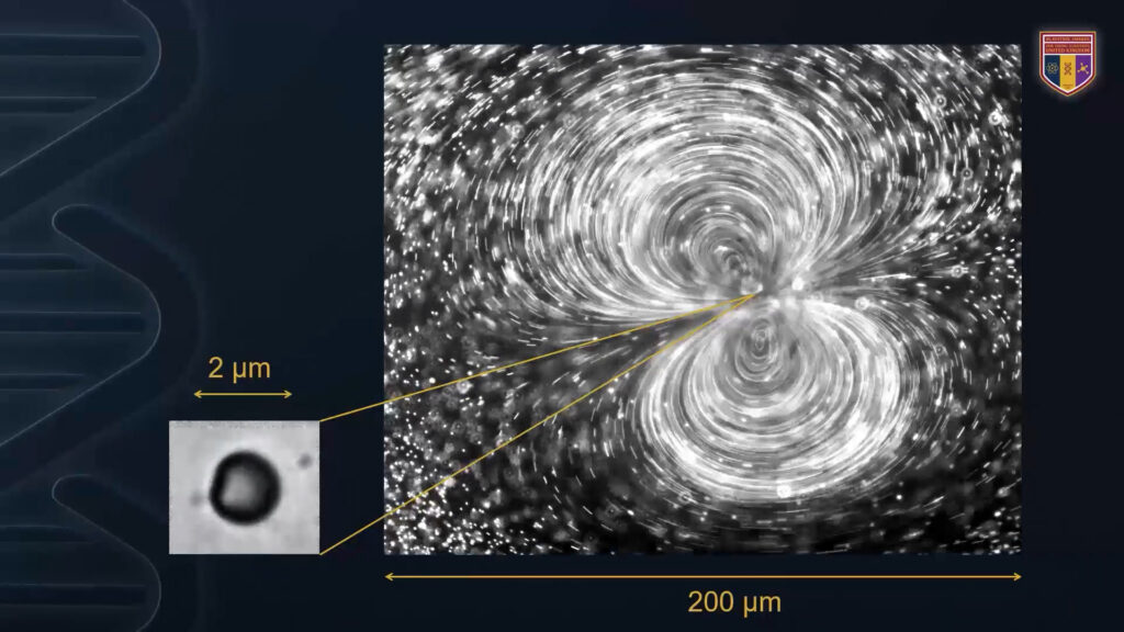
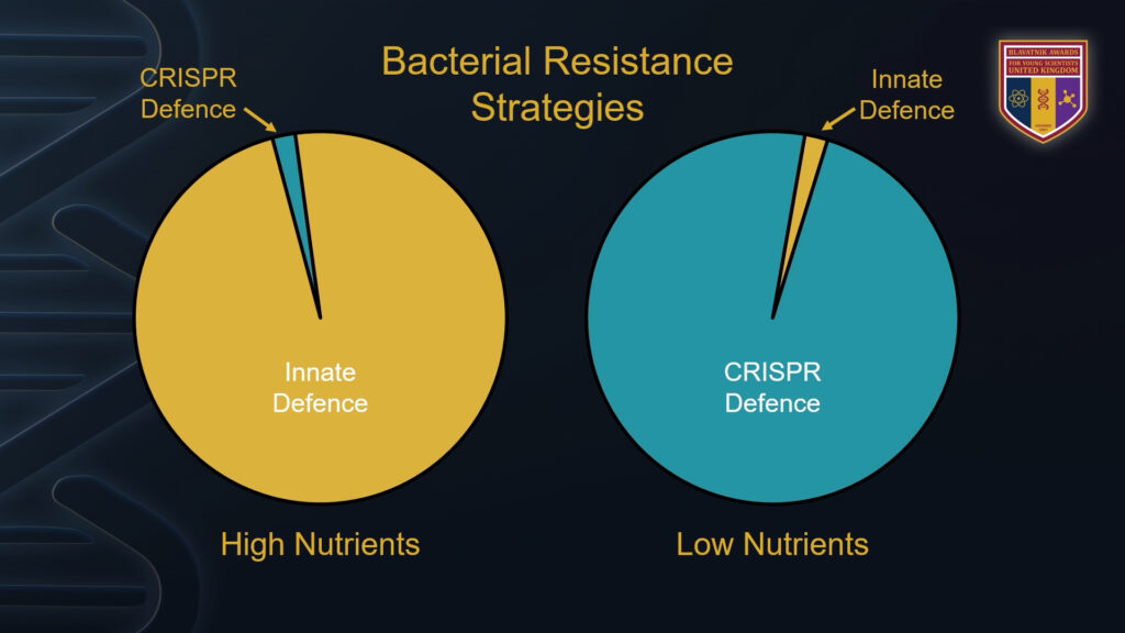

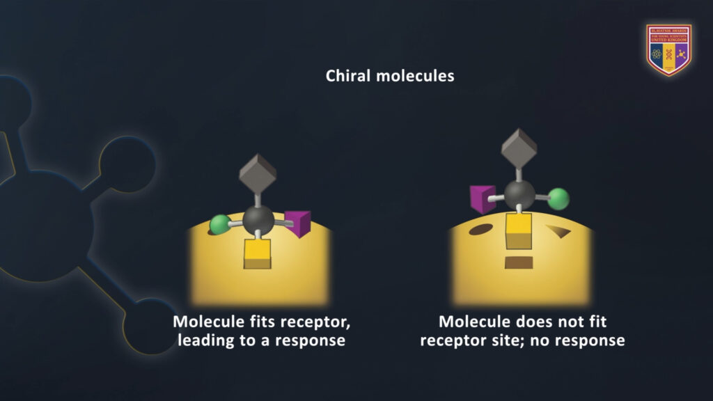
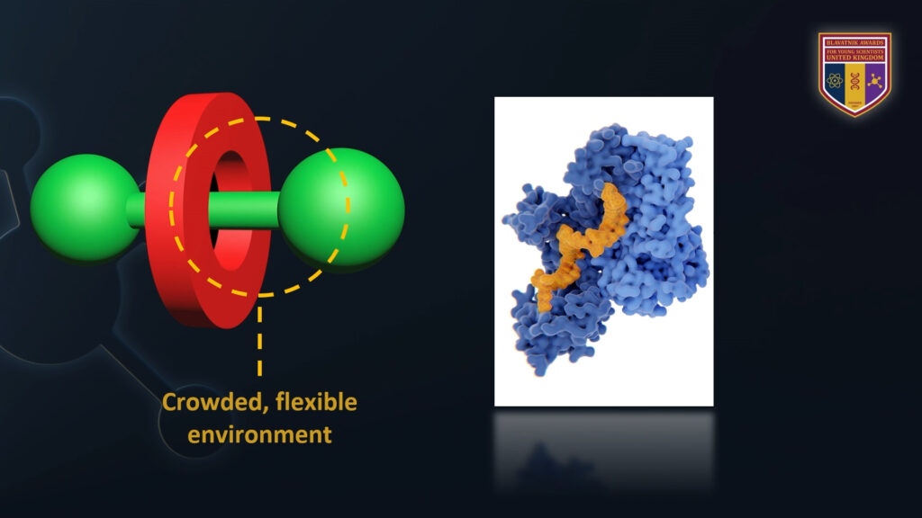
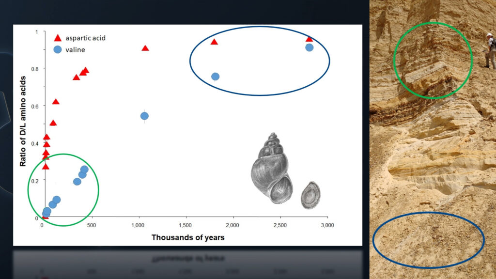
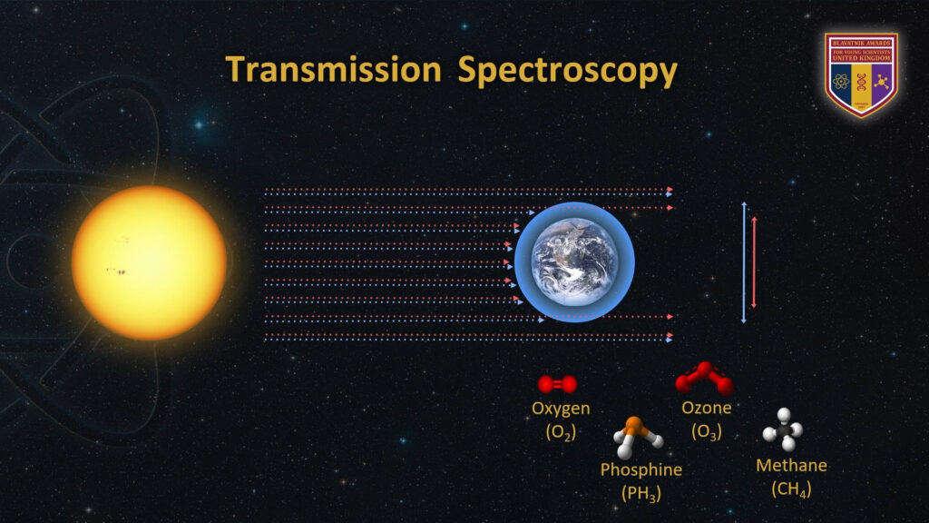
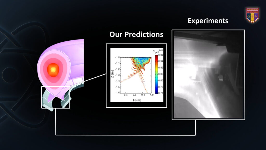
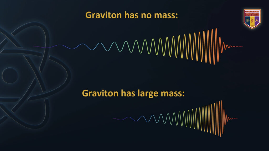


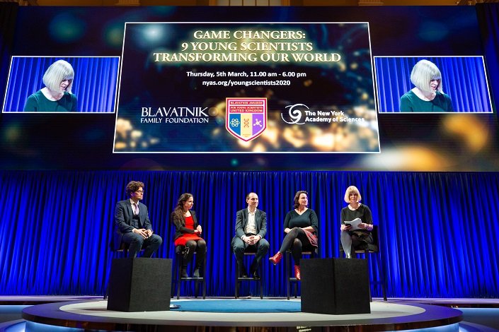
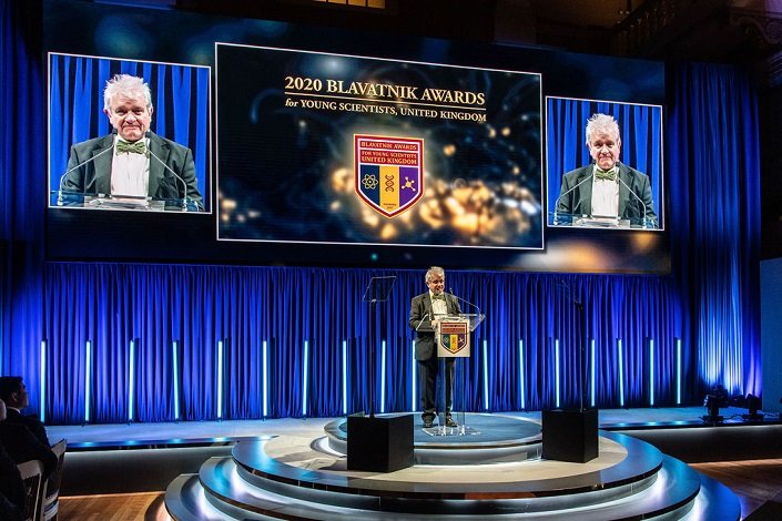

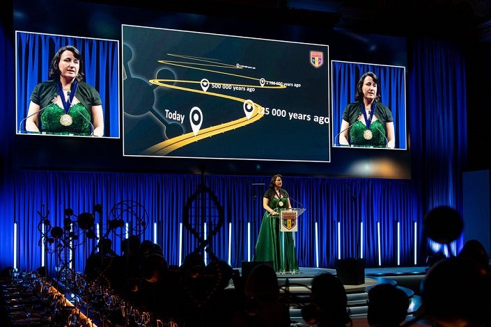


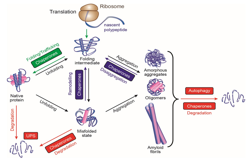
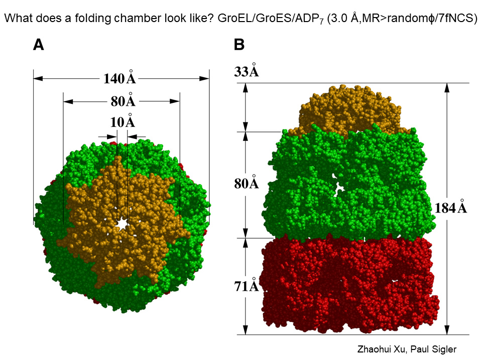
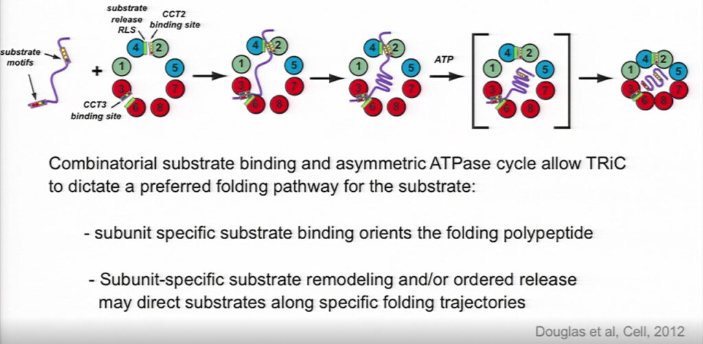
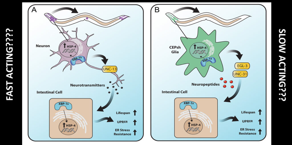
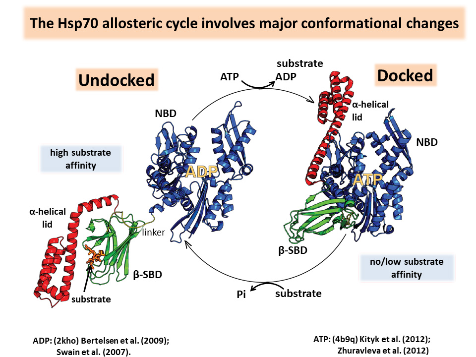
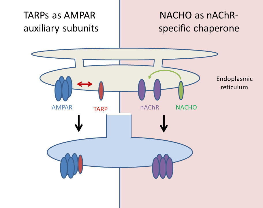
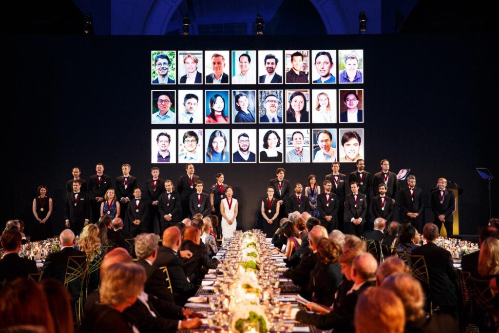
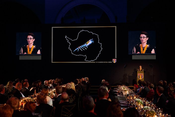
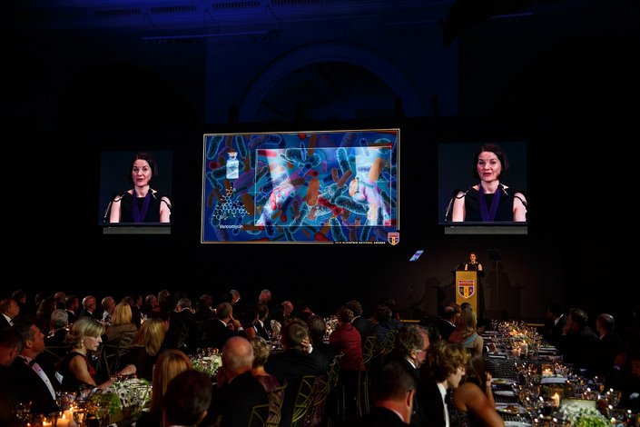
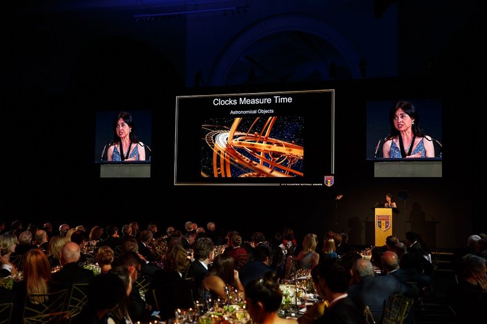


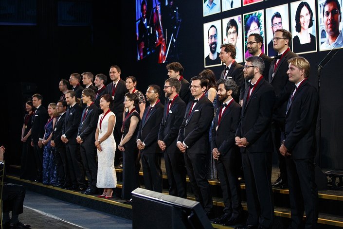
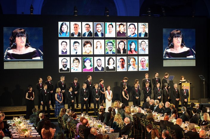
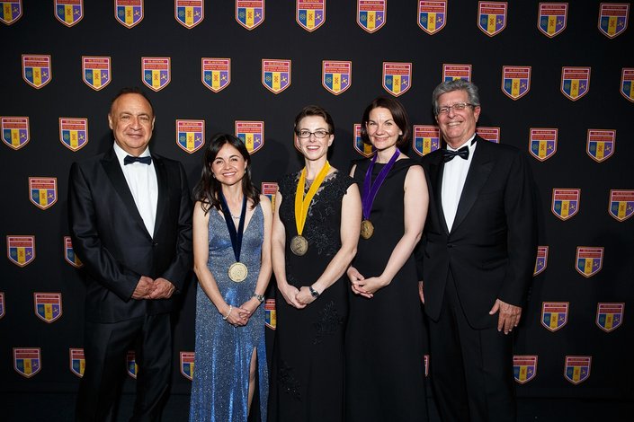


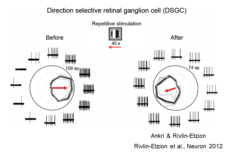
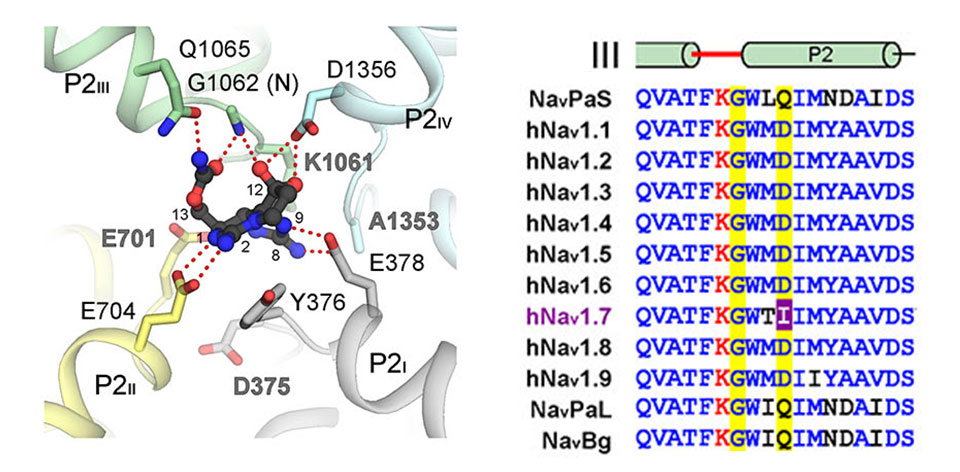
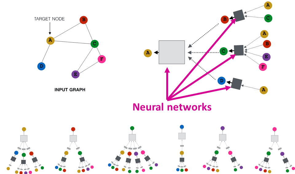
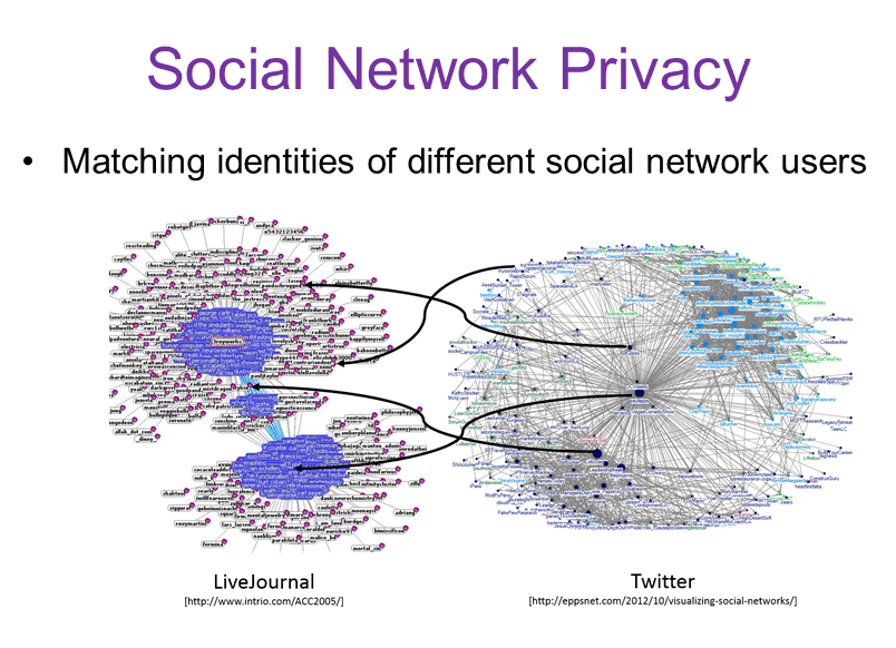


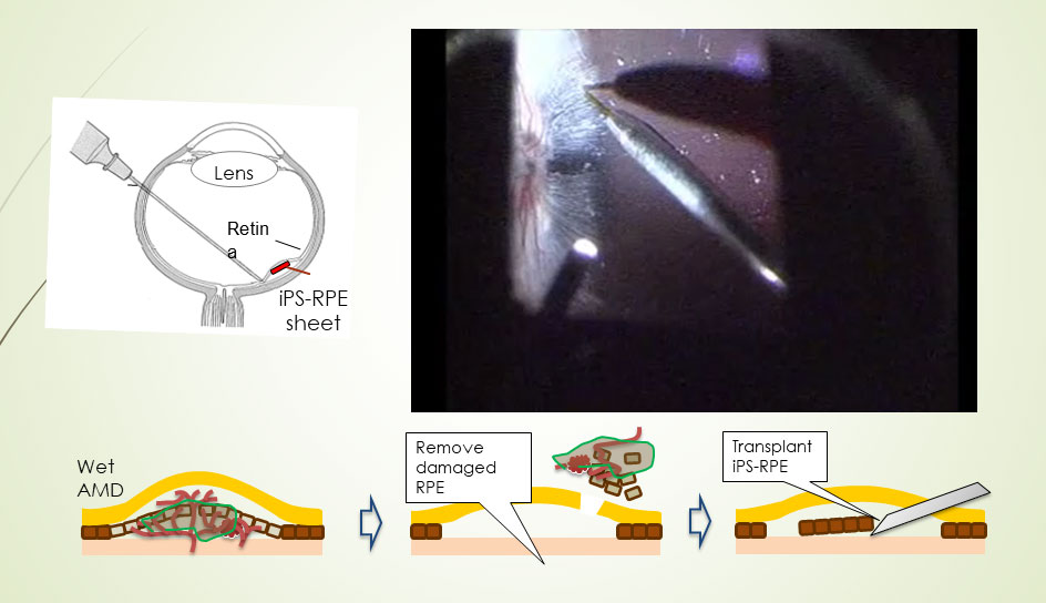
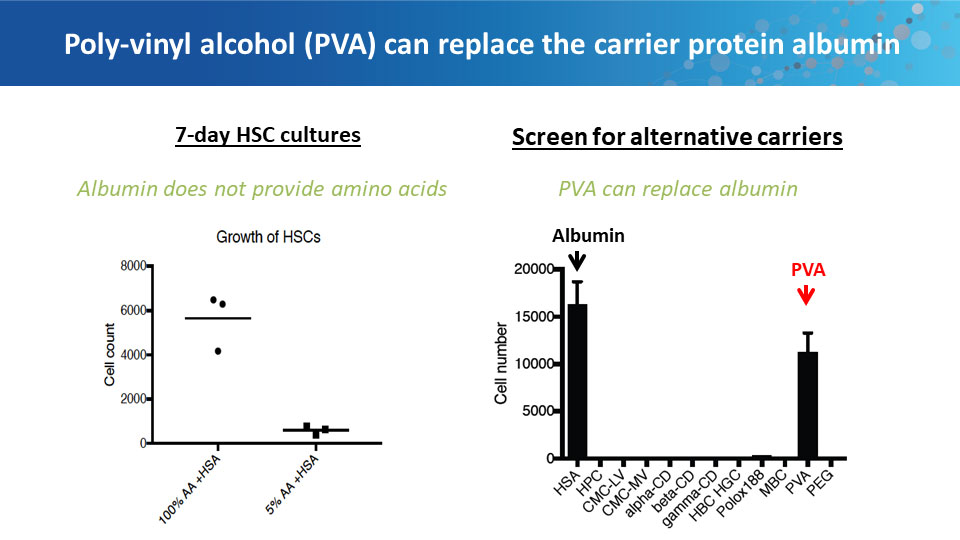
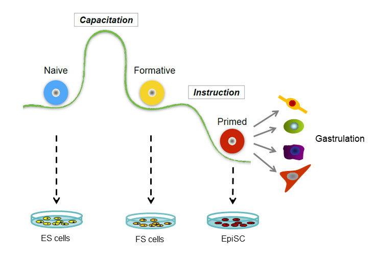
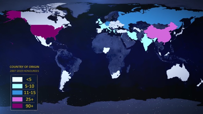


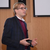
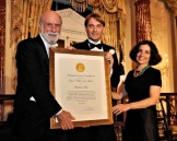
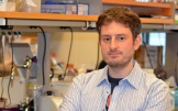